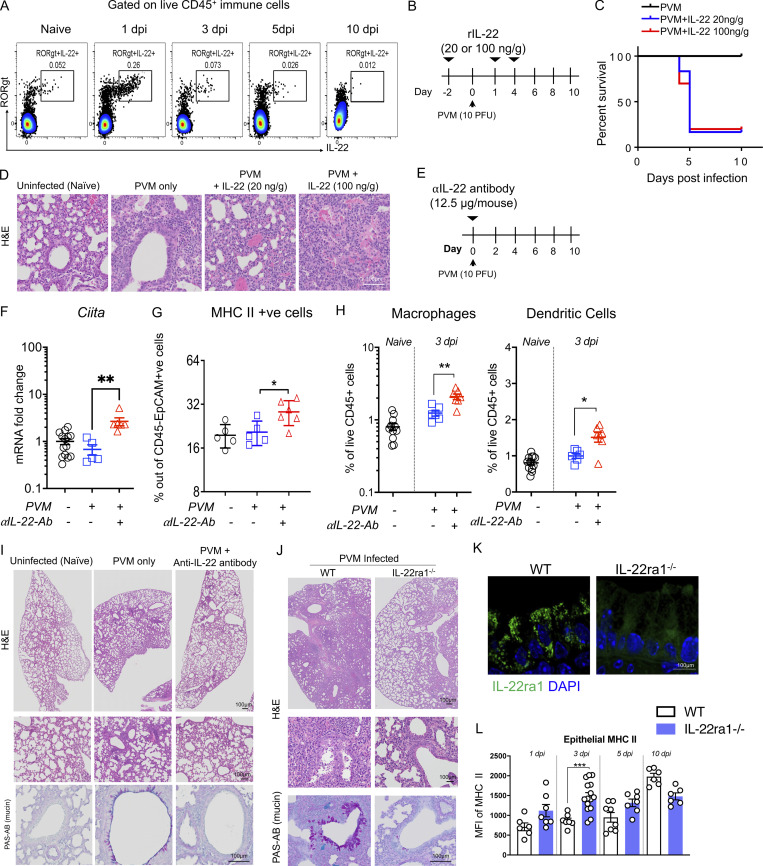
Figure 4.
IL-22 suppression of epithelial MHC II is associated with exacerbated pathology in acute respiratory viral infection. (A) Flow cytometry plots showing abundance of RORγt+ IL-22+ respiratory tract immune cells after infection of C57BL/6 mice with 10 PFU PVM at indicated dpi. (B) Schematic experimental diagram of rIL-22 (20 or 100 ng/g) treatment prior to PVM infection. (C) Survival curves. (D) Representative H&E-stained lungs collected on day 10. (E) Schematic of experiment with PVM-infected C57BL/6 animals treated with α-IL22-antibody (12.5 μg/mouse) on the day of infection. (F) Respiratory tract mRNA expression of Ciita by qRT-PCR. (G and H) Flow cytometry determination of the relative frequency of MHC II+ and H macrophages and dendritic cells respiratory tract epithelial cells on day 3 after infection. (I) Representative H&E- and PAS-AB–stained respiratory tract sections. (J) Representative respiratory tract H&E- and PAS-AB–stained respiratory tract sections on day 10 of PVM-infected Il22raWT (Il-22ra1fl/fl; littermate controls) and Il22ra1KO (CMVcre × Il-22ra1fl/fl) mice. (K) Immunofluorescence staining demonstrating the lack of IL-22RA1 on epithelial cells from Il22ra1KO animals. (L) MFI of MHC II expression on respiratory tract epithelial cells during the course of PVM infection in Il22raWT and Il22ra1KO mice. Statistics: mean ± SEM (n = 5–12); data are representative of three independent experiments. One-way ANOVA, Bonferroni’s post hoc test; *P < 0.05, **P < 0.01, and ***P < 0.001.

