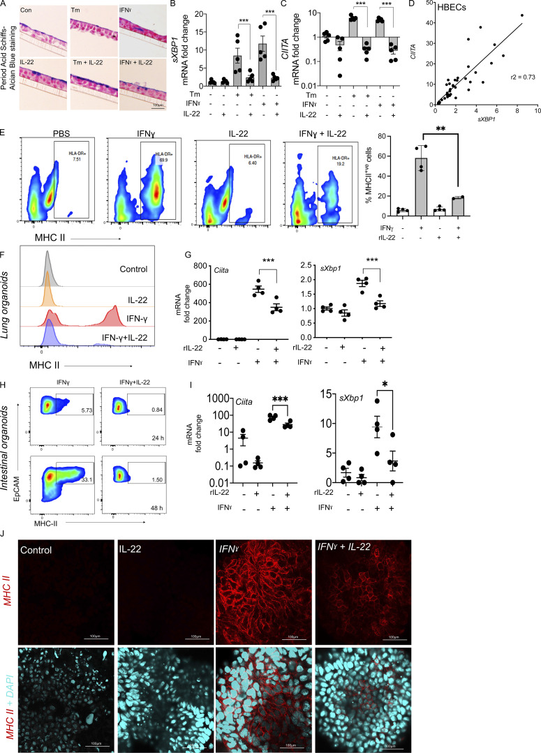
Figure 5.
IL-22 suppresses epithelial MHC II accompanied by a suppression of ER stress. (A) Histological sections of monolayer cultures showing mucin production by HBECs (five donors) treated with Tm (1 µg/ml) or IFNγ (0.1 µg/ml) alone or in combination with IL-22 (10 ng/ml) for 24 h. (B and C) Relative mRNA expression of ER stress marker (sXBP1) and CIITA were measured by qRT-PCR in the HBECs treated as in A. (D) Correlation of CIITA and sXBP1 expression by HBECs. (E) Representative flow cytometric plots and relative frequency of total MHC II expression in HBECs treated with IFNγ (0.1 µg/ml) with and without alone or in combination with IL-22 (10 ng/ml). (F and G) Flow cytometry histograms for cell surface MHC II (F) and (G) qRT-PCR for Ciita and sXbp1 mRNA showing IL-22 (10 ng/ml; 24 h) mediated suppression of IFNγ (1 µg/ml)-induced MHC II and ER stress in cultured murine lung organoids from C57BL/6 mice. (H–J) Flow cytometry scatter plot of intestinal organoids from C57BL/6 mice treated with IFNγ (1 µg/ml) ± IL-22 (10 ng/ml) for 24 or 48 h (H), qRT-PCR of Ciita and sXbp1 mRNA (I), and immunofluorescence staining for MHC II in intestinal organoids (J). Statistics: mean ± SEM (n = 4–12); data are representative of two independent experiments. One-way ANOVA, Bonferroni’s post hoc test; *P < 0.05, **P < 0.01, ***P < 0.001, and ****P < 0.0001.

