Figure S2.
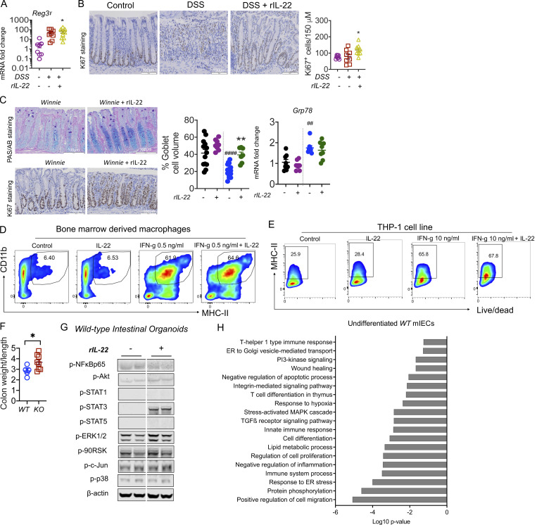
IL-22 reduced pathology during intestinal inflammation. (A) Gene expression level of antimicrobial peptide Reg3γ in the intestine on day 10 of the C57BL/6 mice challenged with 2.5% DSS for the first 6 d and treated with rIL-22 starting from day 3 to day 10. (B) Representative images of Ki67 staining and quantitative data showing Ki67+ve staining increase with rIL-22 treatment in DSS mice. (C) Representative PAS-AB staining and Ki67 staining, blind histological scoring for goblet cell volume, and relative expression of ER stress marker Grp78 in the distal colons of Winnie mice treated with or without rIL-22 on alternative days for 2 wk prior to cull at day 14. (D) Flow cytometric plots and data shown as a % of CD45+, Cd11b+ MHC II+ BMDMs from WT animals in the presence of IFNγ and IL-22 alone or in combination (n = 3). (E) Flow cytometric plots and MHC II+ve cells shown as a percentage of live/dead THP1 cells treated with IFNγ and IL-22 alone or in combination. (F) Colon weight/length ratio of naïve Il22ra1 fl/fl (Il22ra1WT) and CMV-cre × Il-22ra1 fl/fl (Il22ra1KO) mice. (G) Cell signaling activation markers were measured by Western blot analysis in intestinal organoids from WT animals with and without IL-22 (rIL-22, 100 ng/ml for 30 min). (H) DAVID gene ontology analysis of RNA-Seq data shows a downregulation of pathways associated with ER stress, inflammation in WT mice treated with IL-22 (rIL-22, 100 ng/ml for 4 h). Data are representative of three independent experiments and are presented as mean ± SEM (n = 4–12). One-way ANOVA, Bonferroni’s post hoc test; *P < 0.05, **P < 0.01. ##P < 0.01, ####P < 0.0001 compared to untreated WT controls. Source data are available for this figure: SourceData FS2.

