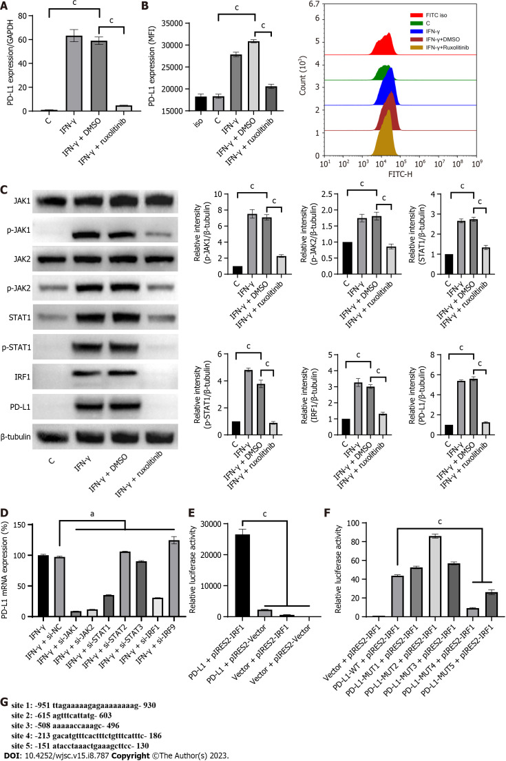
Figure 2.
Interferon-gamma upregulated programmed cell death protein 1 ligand 1 expression via the JAK/STAT1/ interferon regulatory factor 1 pathway in human umbilical-cord-mesenchymal stem cells. A: Programmed cell death protein 1 ligand 1 (PD-L1) expression was quantified by quantitative real-time PCR (qRT-PCR) in human umbilical-cord-mesenchymal stem cells (hUC-MSCs) treated with a combination of IFN-γ with or without the JAK inhibitor ruxolitinib (10 µM) for 4 h; B: PD-L1 expression was quantified by flow cytometry in hUC-MSCs treated with a combination of IFN-γ with or without ruxolitinib (10 M) for 24 h; C: Activation of the JAK/STAT/interferon regulatory factor 1 (IRF1) pathway and PD-L1 expression were analyzed by western blot in hUC-MSCs treated with IFN-γ (20 ng/mL) with or without ruxolitinib (10 M) for 24 h; D: The PD-L1 expression was quantified by qRT-PCR in hUC-MSCs transfected with siRNA against JAK1, JAK2, STAT1, STAT2, STAT3, IRF1, or IRF9 and subsequently treated with IFN-γ for 4 h. The expression of PD-L1 in hUC-MSCs of the IFN-γ + si-NC (negative control) group was used as the standard (100%); E: PGL4.10-PD-L1 promoter, pIRES2-IRF1, and Renilla luciferase plasmids were transfected together or separately into 293T cells. Cells were lysed 24 h after transfection and luciferase activity was measured; F: pIRES2-IRF1 plasmid and Renilla luciferase plasmid were transfected into 293T cells with the PGL4.10-PD-L1 promoter plasmid or IRF1 binding site mutant PGL4.10-PD-L1 promoter plasmids. Cells were lysed 24 h after transfection and luciferase activity was measured; G: Predicted binding sites of IRF1 on the PD-L1 promoter. Data were represented as mean ± SEM of n = 3. C: Control; Iso: Isotype; aP < 0.01; cP < 0.0001. IFN-γ: Interferon-gamma; PD-L1: Programmed cell death protein 1 ligand 1.

