Fig. 5.
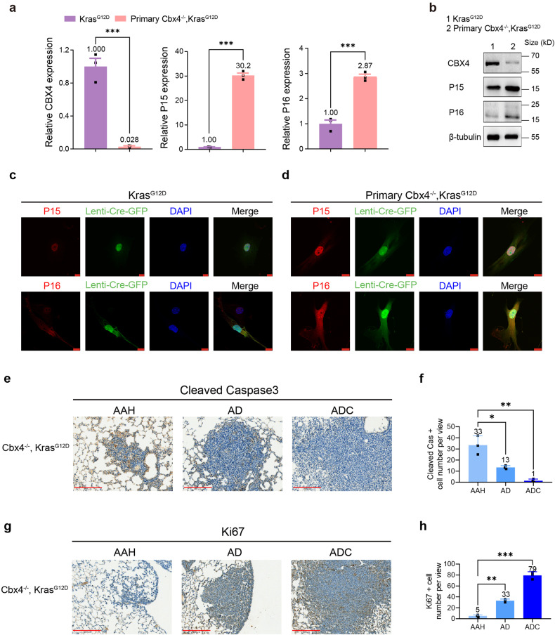
CBX4 loss up-regulates P15, P16 and causes alterations of apoptosis-related genes in primary Cbx4−/−, KrasG12D cells. a Real-time PCR analysis of CBX4, P15 and P16 in KrasG12D and primary Cbx4−/−, KrasG12D MEFs. b Western Blot of CBX4, P15 and P16 in KrasG12D and primary Cbx4−/−, KrasG12D MEFs. c IF staining of P15 (red, up), P16 (red, down), Lenti-Cre-GFP (green) and DAPI (blue) in KrasG12D cells. Scale bar: 10 μm. d IF staining of P15 (red, up), P16 (red, down), Lenti-Cre-GFP (green) and DAPI (blue) in primary Cbx4−/−, KrasG12D cells. Scale bar: 10 μm. e Representative immunohistochemical staining of cleaved Caspase3 in lung sections from Cbx4−/−, KrasG12D mice. Scale bar: 200 μm. f Quantitative analysis of cleaved Caspase3 positive cell number in IHC staining of lung sections of AAH, AD and ADC from Cbx4−/−, KrasG12D mice. g Representative immunohistochemical staining of Ki67 in lung sections from Cbx4−/−, KrasG12D mice. Scale bar: 200 μm. h Quantitative analysis of Ki67 positive cell number in IHC staining of lung sections of AAH, AD and ADC from Cbx4−/−, KrasG12D mice. Data are shown as means ± SEM. *p < 0.05, **p < 0.01, ***p < 0.001

