
Figure 7.
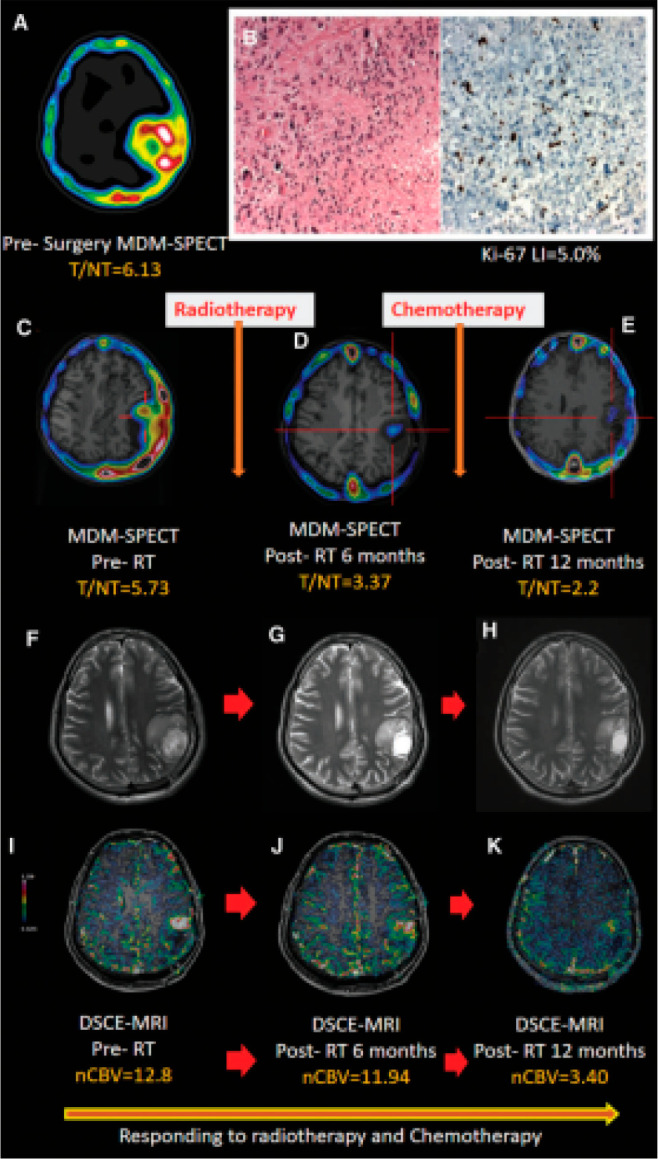
(A) Presurgery imaging of [99mTc]MDM SPECT in a 28-year-old male patient with GBM-IV showing an enlarged tracer uptake (T/NT = 6.13) in the left parietal lobe. (B) Histology (H&E staining, 40×) images show (left panel) several mitoses with regions of palisading necrosis, moderate cellular and nuclear pleomorphism, and lower proliferation index of Ki-67 LI = 5.0% (right panel). In the follow-up scans, serial axial T2-weighted MR images (F, G, and H) did not show any noticeable enhancement on the tumor bed. A significant treatment response (Responder) was shown by follow-up SPECT/MRI, which showed that the baseline pre-radiotherapy (C) tracer uptake (5.73) and (I) nCBV (12.8) decreased (D, J) after 6 months (T/NT = 3.36, nCBV = 11.94) and (E, K) at 12 months (T/NT = 2.3, nCBV = 3.40). Abbreviations: GBM, glioblastoma multiforme; nCBV, normalized cerebral blood volume; SPECT, single-photon emission computed tomography; T/NT, target to non-target; [99mTc]MDM, 99mTc-DTPA-bis(Met). Reproduced with permission from ref (98). Copyright 2021 Mary Ann Liebert, Inc.