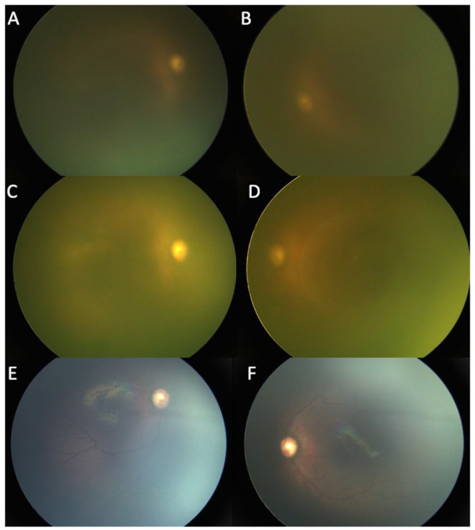
Figure 1.

(A and B) Bilateral fundus photographs in Case 1 at 32 weeks postconceptional age (PCA) show a deteriorating view of the fundus. The total and direct bilirubin levels were 13.3 mg/dL and 11.7 mg/dL, respectively. Fundus photographs of the right eye (C) and left eye (D) in Case 1 at 33 weeks PCA show complete icteric vitreous obstructing fundus visualization. The total and direct bilirubin levels were 17.3 mg/dL and 15.2 mg/dL, respectively. Fundus photographs of the right eye (E) and left eye (F) in Case 1 at 35 weeks PCA show no ROP, no plus disease, and vessels reaching zone 2. The total and direct bilirubin levels were 4.5 mg/dL and 2.5 mg/dL, respectively.
