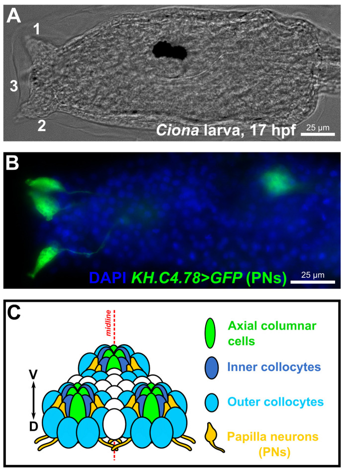
Fig. 1.

The sensory/adhesive papillae of the Ciona larva. (A) Brightfield image of a Ciona robusta (intestinalis type “A”) larva at 17 hpf raised at 20°C, showing the three protruding papillae of the head (numbered 1-3). Papilla number 3, the medial/ventral papilla, is out of focus. (B) Image of electroporated Ciona larva at 17 hpf/20°C, papilla neurons (PNs) labeled by the reporter plasmid KH.C4.78>Unc-76::GFP (green, from Johnson et al., 2023 preprint). Nuclei counterstained by DAPI (blue). (C) Summary diagram of the arrangement and cell type diversity of the papillae (from Johnson et al., 2023 preprint).
