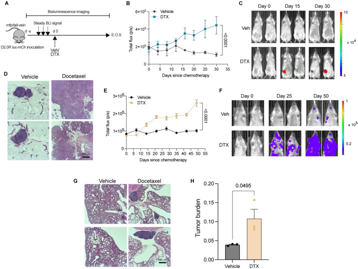
Fig 4. In vivo breast cancer dormancy and docetaxel-mediated dormancy outgrowth.
(A) Schematic showing mouse model of primary (breast) or metastatic (lung) dormancy by injection of D2.0R luc-mCherry cells in the fourth right inguinal mfp or tail-vein, respectively; days since chemotherapy (d), docetaxel 8 mg/kg (DTX), end of study (E.O.S.), and vehicle (Veh). (B, C) Bioluminescence flux kinetics (B) and representative BLI images (C) of D2.0R luc-mCherry tumor growth in the mfp of mice in docetaxel-treated group (n = 9) compared with vehicle-treated controls (n = 7). Two-way mixed ANOVA with post hoc Dunnett’s multiple comparisons test shows statistical significance between treatment groups. (D) Representative gross images of mfps (fourth right inguinal), HE staining are shown from vehicle and docetaxel-treated mice; scale bars, 550 μm. (E, F) Bioluminescence flux kinetics (E) and representative BLI images (F) of D2.0R luc-mCherry tumor growth in the lungs of mice in docetaxel-treated group (n = 5) compared with vehicle-treated controls (n = 4). Two-way mixed ANOVA with post hoc Dunnett’s multiple comparisons test shows statistical significance between treatment groups. (G, H) Representative gross images of mouse lungs (G), HE staining, and tumor burden quantification (H) are shown from vehicle and docetaxel-treated mice; scale bars, 550 μm. Independent t test measurement shows statistical significance between treatment groups. Source data can be found in S1 Data. DTX, docetaxel; HE, hematoxylin and eosin; mfp, mammary fat pad.

