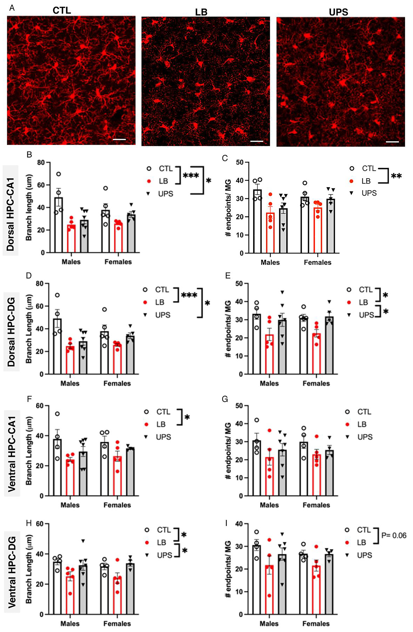
Fig. 2.

Greater reduction in microglial ramification is seen in LB compared to UPS. (A) representative confocal images of Iba1 staining in the dorsal hippocampus of P17 pups exposed to CTL, LB, and UPS. Effects of rearing and sex on microglial branch length (B, D, F, H) and number of branch-endpoints (C, E, G, I) in the dorsal hippocampus CA1 region (B-C), dorsal hippocampus dentate gyrus (D-E), ventral hippocampus CA1 region (F-G), ventral hippocampus dentate gyrus (H-I). N = 4–7 mice per rearing and sex group. Scale bars in A are 20 μm. Error bars represent mean ± SEM. *p < 0.05, **p < 0.01, ***p < 0.001.
