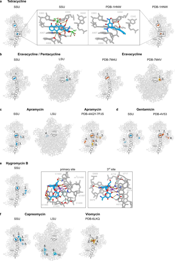
Extended Data Fig. 5. Secondary binding sites of antibiotics on the ribosome.
a, Overview of the primary (1.) and secondary (2.) binding sites of tetracycline (blue) on the SSU determined here (left) and tetracycline (red) reported previously on the SSU (right, PDB ID 1HNW)26, with boxed zooms revealing the inverse orientation of tetracycline and distinct interactions observed with the rRNA. b, primary (1.) and secondary (2.-3.) binding sites of eravacycline/pentacycline (blue/cyan) on the SSU and LSU (left), compared with sites (red/orange) reported previously for eravacycline on the LSU (right)(PDB ID 7M4V)31. c, primary (1.) and secondary (2.-4.) binding sites of apramycin (blue) on the SSU and LSU (left), compared with sites reported previously on the LSU (right)(PDB ID 4AQY and 7PJS)7,29. d, primary (1.) and secondary (2.-3.) binding sites of gentamicin (blue) on the SSU (right), compared with sites reported previously for gentamicin (red/orange) on the SSU (right)(PDB ID 4V53))28. e, primary (1.) and secondary (2.-3.) binding sites of hygromycin B (blue) on the SSU (left), with insets showing the similarity in the coordination of a putative K+ ion in the primary and 3rd site. f, primary (1.) and secondary (2.-4.) binding sites of capreomycin (blue) on the SSU and LSU (left), compared with sites reported previously for viomycin on the SSU (right)(PDB ID 6LKQ)33.

