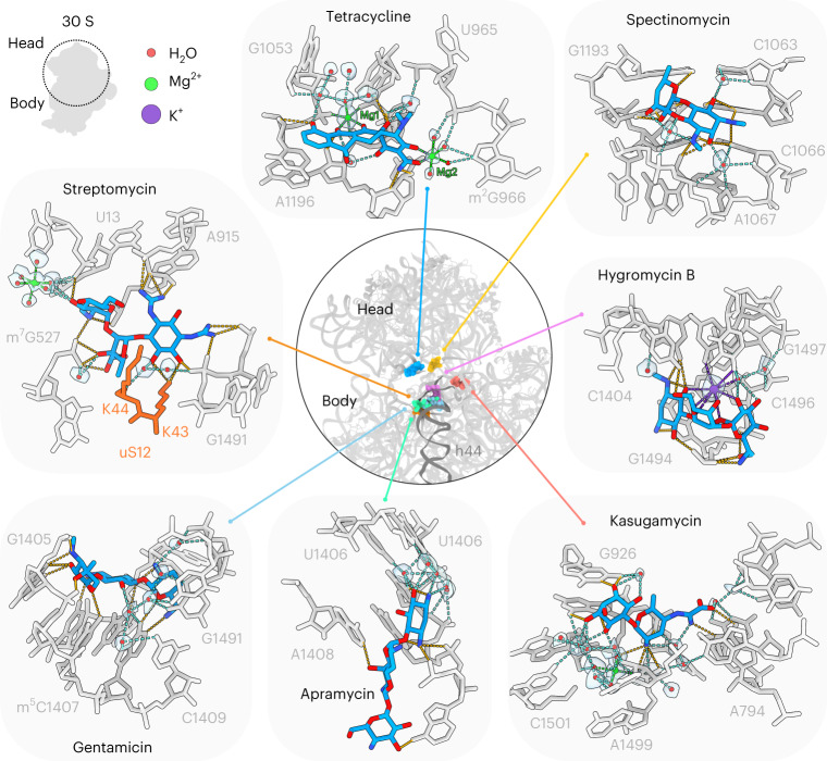
Fig. 2. Structures of antibiotics targeting the SSU.
The central ring shows a superimposition of the binding sites on the SSU (gray) of the antibiotics tetracycline (blue), spectinomycin (yellow), hygromycin B (pink), kasugamycin (red), apramycin (green), gentamicin (cyan) and streptomycin (orange), which is surrounded by insets highlighting the interactions between the drug and the 16S rRNA (gray), waters (red spheres with gray transparent density), magnesium ions (green spheres), putative K+ ions (purple sphere with transparent gray density) and uS12 (orange). Potential hydrogen bonds are indicated as dashed lines, colored orange for direct interaction between the drug and the small subunit, cyan for water-mediated interactions, green for Mg2+ ion coordination and purple for K+ coordination.

