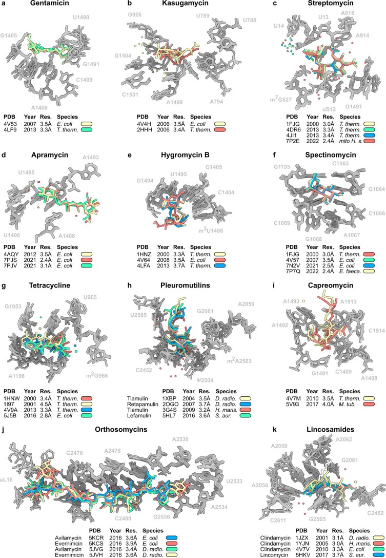
Extended Data Fig. 1. Comparison of structures of antibiotic-ribosome complexes.
a-k, Superimposition of previous structures of diverse antibiotic-ribosome complexes, including (a) gentamicin (yellow) on the E. coli 70S ribosome at 3.5 Å (PDB ID 4V53)28 with gentamicin (green) on the T. thermophilus 30S at 3.3 Å (PDB ID 4LF9), (b) kasugamycin (yellow) on the E. coli 70S ribosome at 3.5 Å (PDB ID 4V4H)89 with kasugamycin (red) on the T. thermophilus 30S at 3.4 Å (PDB ID 2HHH)30, (c) streptomycin on the T. thermophilus 30S at 3.0 Å (yellow; PDB ID 1FJG)90 at 3.3 Å (green, PDB ID 4DR6)91 at 3.35 Å (blue, PDB ID 4JI1)91 with streptomycin (red) on the human mitochondrial small subunit at 2.4 Å (PDB ID 7P2E)92, (d) apramycin (yellow) on the T. thermophilus 30S at 3.5 Å (PDB ID 4AQY)29 with apramycin on the E. coli 70S ribosome at 2.4 Å (red, PDB ID 7PJS)7 and 3.1 Å (green, PDB ID 7PJV)7, (e) hygromycin B on the T. thermophilus 30S at 3.3 Å (yellow, PDB ID 1HNZ)26 and 3.7 Å (blue, PDB ID 4LFA) with hygromycin B on the E. coli 70S ribosome at 3.5 Å (red, PDB ID 4V64)37, (f) spectinomycin (red) on the T. thermophilus 30S at 3.0 Å (PDB ID 1FJG)90 with spectinomycin on E. coli 70S ribosome at 3.5 Å (green, PDB ID 4V57)28, E. faecalis 70S ribosome at 2.4 Å (yellow, PDB ID 7P7Q)8 and within an E. coli 70S translocation intermediate at 2.5 Å (blue, PDB ID 7N2V)55, (g) tetracycline on the T. thermophilus 30S at 3.4 Å (red, PDB ID 1HNW)26 and 4.5 Å (yellow, PDB ID 1I97)27 with tetracycline (blue) on the T. thermophilus 70S at 3.3 Å (PDB ID 4V9A)93 and tetracycline (green) on the E. coli 70S at 2.8 Å (PDB ID 5J5B)94, (h) the pleuromutilins tiamulin (yellow) on the D. radiodurans 50S at 3.5 Å (PDB ID 1XBP)50, retapamulin (blue) on the D. radiodurans 50S at 3.7 Å (PDB ID 2OGO)51, tiamulin (red) on the archaeal H. marismortui 50S at 3.2 Å (PDB ID 3G4S)49 and lefamulin (green) on the S. aureus 50S at 3.6 Å (PDB ID 5HL7)53, (i) capreomycin (yellow) on the T. thermophilus 70S at 3.5 Å (PDB ID 4V7M)32 with capreomycin (red) on the M. tuberculosis 70S at 4.0 Å (PDB ID 5V93)95, (j) the orthosomycins avilamycin (blue, PDB ID 5KCR) and evernimicin (red, PDB ID 5KCS) on the E. coli 70S at 3.9 Å43 with avilamycin (green, PDB ID 5JVG) and evernimicin (yellow, PDB ID 5JVH) on the D. radiodurans 50S at at 3.6 Å and 3.4 Å, respectively44, (k) the lincosamide clindamycin on the D. radiodurans 50S at 3.1 Å (yellow, PDB ID 1JZY)96, on the H. marismortui 50S at 3.0 Å (red, PDB ID 1YJN)97 and on the E. coli 70S at 3.3 Å (green, PDB ID 4V7V)98 with lincomycin on the S. aureus 50S at 3.7 Å (blue, PDB ID 5HKV)99. Alignments were made using the rRNA within 10 Å of the antibiotic. rRNA and r-proteins comprising the binding site are colored grey, whereas antibiotics (including waters and ions if present) are color-coded as indicated.

