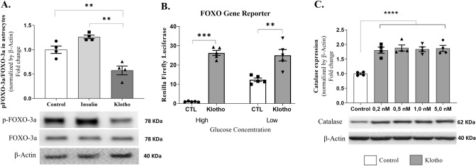
Figure 2.
Klotho affects FOXO-3a activity and catalase activity in astrocytes. (A) Western blot assay was performed to measure phosphorylation levels of FOXO-3a. Statistical analysis suggested that Klotho treatment decreased the FOXO-3a inhibitory phosphorylation. One way ANOVA followed by Tukey's multiple comparisons tests; [F = 23.86; P = 0.0003; R squared = 0.8413]. Below the graph is the representative western blot digital images of p-FOXO-3a, FOXO-3a, and β-actin. (B) FOXO gene reporter activity. After 48 h of plasmid transfections, treatments were performed by incubating cells for 24 h with DMEM containing high (4.5 g/L) or low (1 g/L) concentrations of glucose, with or without 1 nM Klotho. Results represent the ratio of fluorescence of firefly luciferase to renilla luciferase, normalized to the mean of the control group (DMEM high glucose). One-way ANOVA statistical analysis followed by Tukey’s multiple comparison tests. FOXO-3a activity was increased in all groups: CTL High vs. CTL Low (mean difference: − 11.35; q = 6.782), CTL High versus Klotho High (mean difference: − 25.26; q = 15.09), CTL High versus Klotho Low (mean difference: − 24.06; q = 14.38), CTL Low versus Klotho High (mean difference: − 13.91; q = 8.313), and CTL Low versus Klotho Low (mean difference: − 12.71; q = 7.598). No difference was observed between Klotho high and Klotho low glucose (mean difference: 1.196; q: 0.7148). P < 0.001, F = 50.66, and R squared = 0.9048. (C) Catalase relative activity. Statistical analysis suggested that all klotho concentrations increased catalase expression compared to that of the control group. [One-way ANOVA followed by Tukey’s multiple comparison test. F = 18.38; R squared 0.8386; P < 0.0001]. *P < 0.05; ***P < 0.0001. The original autoradiographs are available in Supplementary Figs. S4 and S5. Lysates were obtained from two or three wells of a 6-well plate to reach 2 × 10⁶ cells. Each point in the graphs represents a lysate obtained from independent cell cultures, and four independent cell cultures were analyzed in these assays.

