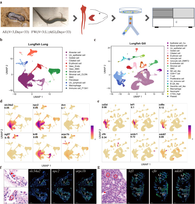
Fig. 1. Single-cell transcriptional profiles of West African lungfish lung and gill under freshwater and terrestrialized conditions.
a Experimental workflow in this study. AE denotes terrestrialization; FW, freshwater. L, lung; G, gill. b Lung cell landscape of 53,605 cells from terrestrialized and freshwater lungfish. Cells are shown by Uniform Manifold Approximation and Projection and color-coded by cluster cell type. Each dot represents a cell, and different colors are associated with specific cell types. Vasc_Endo, vascular endothelial cell; Vasc_SMC, vascular smooth muscle cell; SMC, smooth muscle cell; Un_epithelial cell, unclassified epithelial cell; Un_lymphoid cell, unclassified lymphoid cell. c Gill cell landscape of 87,347 cells from terrestrialized and freshwater lungfish. Cells are shown by UMAP and color-coded by cluster cell type. Each dot represents a cell, and different colors are associated with specific cell types. NK cell_like, Natural killer cell_like; Un_epithelial cell, unclassified epithelial cell; Un_immune cell, unclassified immune cell. d Cell type identification of six classical lung cell type markers (see Supplementary Fig. 1c for overall markers). Markers are shown by density plots. Values indicate the max density. The color scale is relative, ranging from 0 to highest density. e Cell type identification of six classical gill cell type markers (see Supplementary Fig. 1d for overall markers). Annotated as in (d). f Confirmation of scRNA-Seq cell type annotation results by H&E stanning and fluorescence microscopy image of African lungfish lung. Green, digoxigenin-labeled marker genes probes amplified using FITC-TSA; blue, DAPI (scale bar = 500 μm). (1) fibromuscular wall; (2), air sacs. On the H&E image, the red solid circle represents alveolar epithelial cells; red rhombus represents macrophage cells; red circle represents the lymphoid nodes. Each slide was repeated independently 3–5 times. Scalebar, 500μm. g Confirmation of scRNA-Seq cell type annotation results by H&E stanning and fluorescence microscopy image of African lungfish gill. Annotated as in (f). Fl, filament; Cr, cartilage; Wight arrows, lamella. Each slide was repeated independently 3–5 times.

