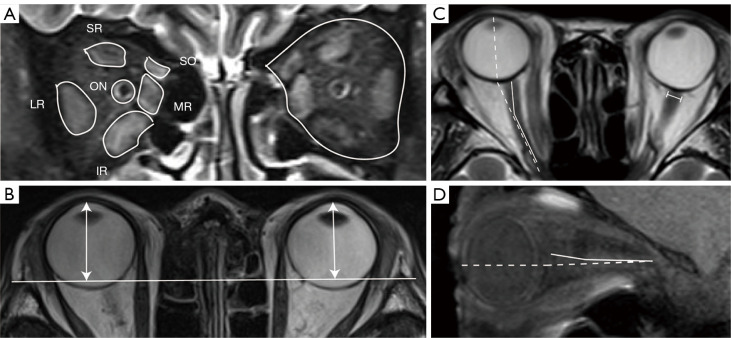
Figure 1.
Measurements of orbital MRI data on T2-weighted images. (A) Measurements of the cross-sectional area of whole orbit and EOMs, including the SR, IR, LR, MR, SO, and ON. (B) Measurement of proptosis. The perpendicular distance (dual arrows) from the top of the cornea to the line connected to bilateral frontal processes in the zygomatic bones was measured. (C) Measurements of the axial ONA (solid line), horizontal gaze angle (dotted line), and the diameter of optic nerve sheath (I-shaped line). (D) Measurements of the sagittal ONA (solid line) and vertical gaze angle (dotted line). SR, superior rectus; IR, inferior rectus; LR, lateral rectus; MR, medial rectus; SO, superior oblique; ON, optic nerve; MRI, magnetic resonance imaging; ONA, optic nerve angle; EOM, extraocular muscle.

