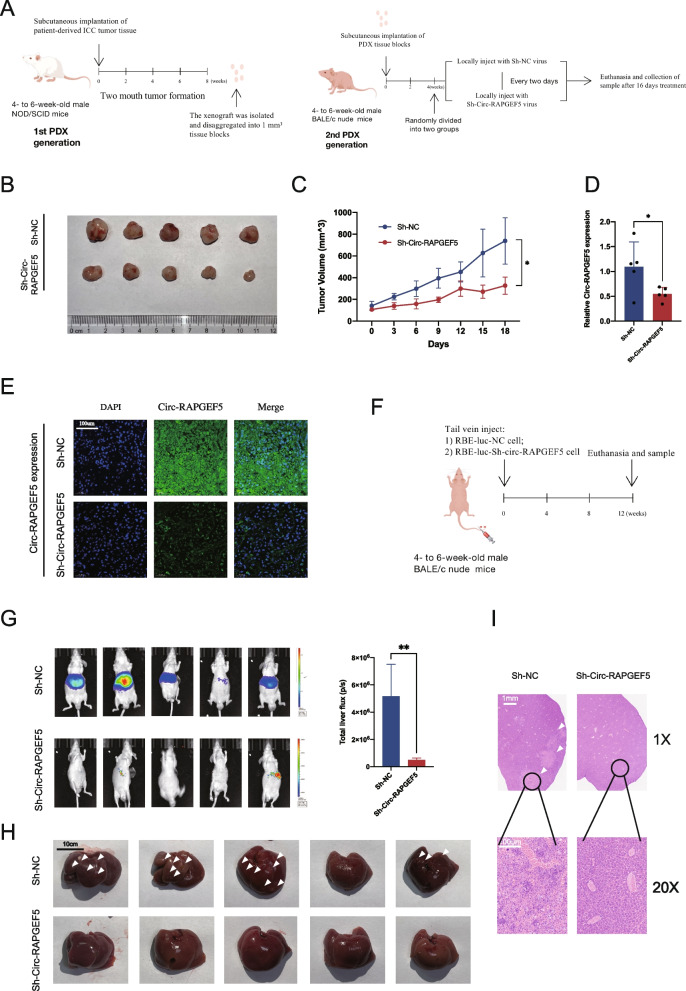
Fig. 3.
Effects of Circ-RAPGEF5 on the proliferation and metastasis in vivo. A NOD/SCID mice (4- to 6-week-old males) were subcutaneously implanted with ICC patient-derived xenograft (PDX). 2 months later, the xenograft was removed for a second round of axillae implantation. On week 4, the second-generation PDX mice were injected at the implantation site within Sh-Circ-RAPGEF5 or Sh-NC lentivirus every two days for 16 days. Animals were sacrificed, and the second-generation PDX was isolated on day 16. B Collection of subcutaneous xenografts of two groups. C Growth curve of xenografts in vivo measured every two days. D qRT-PCR analysis of the relative expression levels of Circ-RAPGEF5 in xenografts tissue of two groups. E The expression of Circ-RAPGEF5 in two groups detected by FISH (20x). F Liver metastasis models were established by injecting Sh-NC and Sh-Circ-RAPGEF5 RBE-luc cells via the tail vein. G Representative bioluminescent images and comparison of fluorescence intensity of liver metastasis. H Image of liver metastasis in two groups. The arrow indicates the metastatic lesions. I Representative H&E staining of liver metastatic lesions. All data are presented as the means ± SD. *p < 0.05, **p < 0.01, ***p < 0.001, ****p < 0.0001

