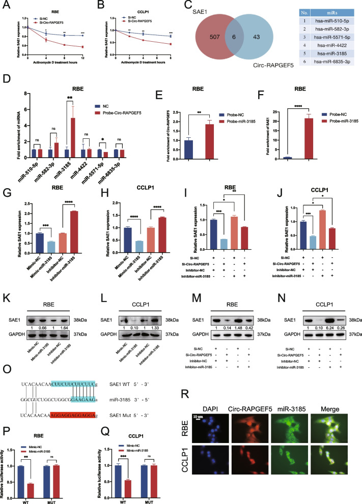
Fig. 7.
Circ-RAPGEF5 competitively sponges miR-3185 to relieve the degradation of miR-3185 on SAE1. A-B qRT-PCR analysis detected relative SAE1 expression in RBE and CCLP1 cells transfected with mock or Si-Circ-RAPGEF5 and treated with Actinomycin D at the indicated time points. C Venn plot illustrating the overlap of predicted 49 microRNAs that could be sponged by Circ-RAPGEF5 and predicted 513 microRNAs that potentially bind to the 3' UTR of SAE1. 6 microRNAs were identified. D RNA pull-down assay using Circ-RAPGEF5-biotin probe revealed that miR-3185 directly interact with Circ-RAPGEF5 in RBE. E–F RNA pull-down assay using a miR-3185-biotin probe confirmed the direct interaction of miR-3185 with Circ-RAPGEF5 (E) and SAE1 (F). G-H qRT-PCR analysis testing SAE1 mRNA expression in knockdown and overexpression of miR-3185 in RBE and CCLP1 cells. I-J qRT-PCR analysis detecting SAE1 mRNA expression in RBE and CCLP1 cells transfected with Si-NC or Si-Circ-RAPGEF5 and inhibitor-NC or inhibitor-miR-3185. K-L Western blot analysis detecting SAE1 protein levels in knockdown and overexpression of miR-3185 of RBE and CCLP1 cells. M–N Western blot analysis detecting SAE1 protein levels in RBE and CCLP1 cells transfected with Si-NC or Si-Circ-RAPGEF5 and inhibitor-NC or inhibitor-miR-3185. O Schematic illustration showed the alignment of miR-3185 with SAE1 3' UTR (blue) and the mutant nucleotides (red). P-Q Dual luciferase reporter assays showed the luciferase activity of wild-type or mutant SAE1 following co-transfection with miR-486-5p mimic or control mimic in RBE and CCLP1 cells. Relative firefly luciferase expression was normalized to that of Renilla luciferase. R FISH assays indicated that Circ-RAPGEF5 and miR-3185 co-localized in the cytoplasm. Circ-RPAGEF5 (red), miR-3185 (green), nuclei staining (blue), and merged (yellow) images in RBE and CCLP1 cells. *p < 0.05, **p < 0.01, ***p < 0.001, ****p < 0.0001

