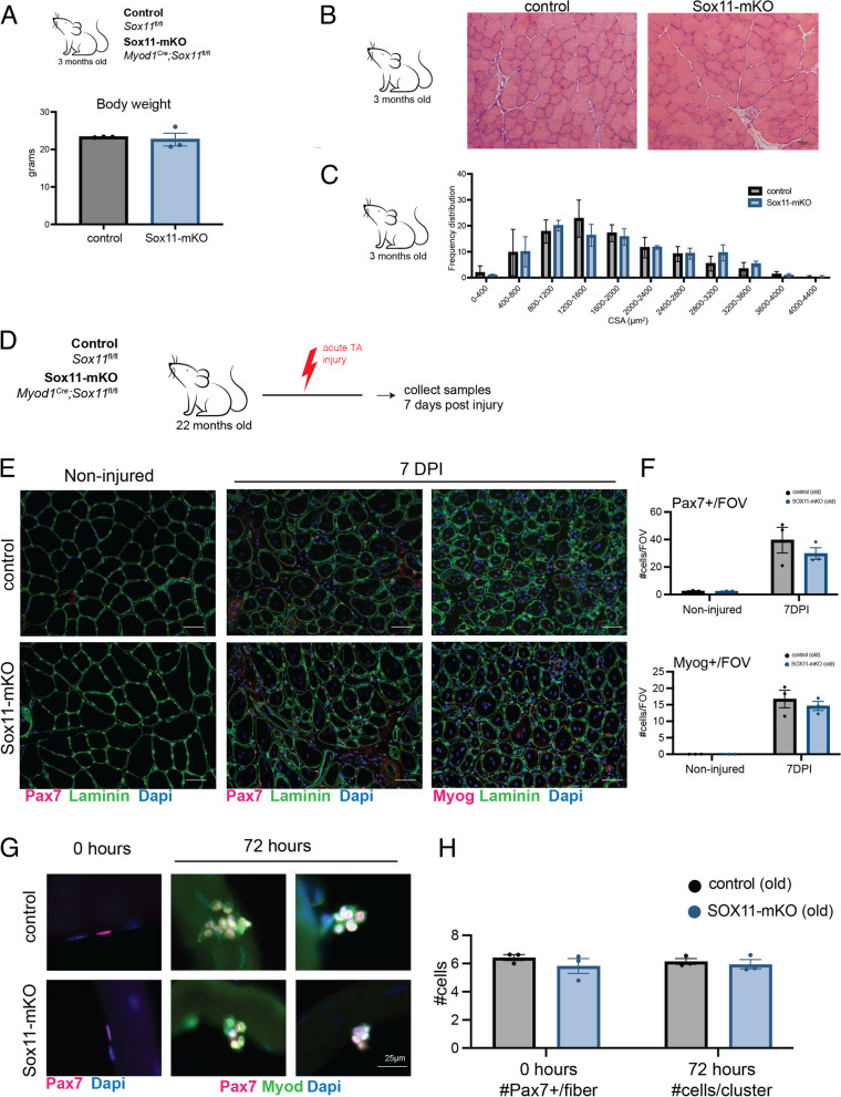
Fig. 6.
Requirement of Sox11 in muscle development and aging. A Body weight of control and Sox11-mKO mice in grams. B H&E staining and representative images of 10-µm TA muscle cross-sections from control (top panel) and Sox11-mKO (bottom panel) non-injured samples, scale bars: 50 µm. C TA muscle fiber CSA for non-injured control and Sox11-mKO mice, related to B. D Experimental outline to evaluate the impact loss of Sox11 has on regeneration of aged (> 20-month-old) mice. The TA muscle of control and Sox11-mKO mice were injured via intramuscular injection of CTX and collected at 7 DPI to evaluate regeneration. E Representative images of immunofluorescence on TA muscle sections to detect Pax7 or Myog, dystrophin, and nuclei (counterstained with DAPI) from control (top panel) and Sox11-mKO (bottom panel) old mice at 0 (non-injured) and 7 DPI, scale bars: 50 µm. F Quantification of the number of Pax7 + cells/FOV (top graph) and Myog + cells/FOV (bottom graph) for control and Sox11-pKO mice. G Ex vivo culture of muscle fibers isolated from EDL of old control (top panel) and Sox11-mKO (bottom) mice, fixed at 0 h (left panel) or after 72 h in culture (right panel) and stained to detect Pax7, Myod, and nuclei (DAPI), scale bar 25 µm. H Quantification of the number of Pax7 + cells/myofiber at 0 h (left graph) and the number of cells/clusters at 72 h in culture (right graph), related to G

