
Figure 3.
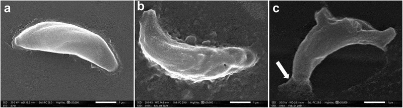
Scanning electron microscopic (SEM) images of.
a. Uniformly crescentic normal, non-treated tachyzoite with a rounded pole and a nearly pointed pole. The site of the conoid was marked at the pointed side by a compressed spring (x20000).
b. CMX-treated tachyzoite showing surface irregularities with multiple depressions and protrusions (x20000).
c. CMX-treated tachyzoite showing completely disrupted morphology with the formation of a dolphin-like structure formed by the combination of deep erosions, several large protrusions, and leakage of internal contents from one end (arrow). The site of the conoid was unremarkable (x20000).