
Figure 5.
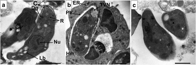
Transmission electron microscopic (TEM) images of.
a. Longitudinal section of a normal, non-treated crescent-shaped extracellular tachyzoite having intact plasma membrane with intact apical complex with conoid (C), rhoptries (R). Dense granules (Dg) and lipid bodies (Lb) were present. The nucleus contains two nuclei with an interrupted nuclear membrane denoting nuclear division (x8000).
b. Longitudinal section of a normal, non-treated crescent-shaped intracellular tachyzoite with host mitochondria (Hm) and endoplasmic reticulum (ER) in close association with the parasitophorous vacuole (PV). Intact tubulovesicular network (TVN) inside the narrow-spaced PV (x6000).
c. CMX-treated tachyzoite showing hazy cytoplasmic membrane, irregular fading nuclear membrane, and disrupted apical complex (x8000).