
Figure 6.
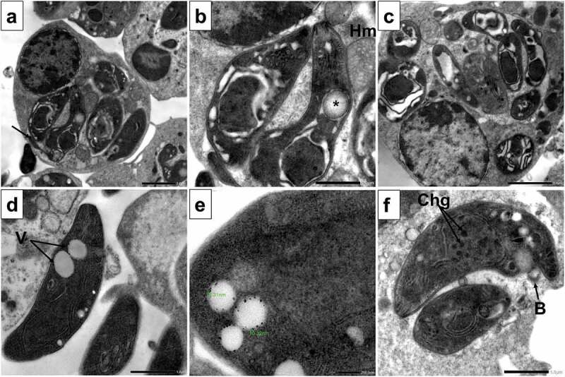
TEM images of SeNPs treated tachyzoites showing.
a. Destabilization of PV membrane with leakage of its contents into the cytoplasm of the host cell. Disrupted tachyzoites morphology with vacuolated cytoplasm and flagella-like membrane protrusion (arrow) (x3000).
b. With higher magnification, the degenerated nuclear membrane of tachyzoites with absence of nucleolus and halo mark around the nucleus were evident. Also disrupted apical complex and appearance of autophagy vesicles containing materials in different stages of degradation were seen (asterisk). tachyzoites showed extensive clefts and close association with host mitochondria (Hm) (x8000).
c. Disruption of PV membrane with leakage of its content into the cytoplasm of the host cell. Multiple tachyzoites showing signs of degradation with extensive cytoplasmic clefts and vacuoles (x4000).
d. Extracellular tachyzoites with multiple vacuoles (V) containing nanosized particles (x8000).
e. With higher magnification, the particle size is between 10.32 and 16.31 nm (x20000).
f. Tachyzoite showing membrane blebs (B), disrupted nuclear membrane with multiple dense chromatin granules (Chg) inside the nucleus. The PV space was widened with disintegrated contents (x8000).