Figure 7.
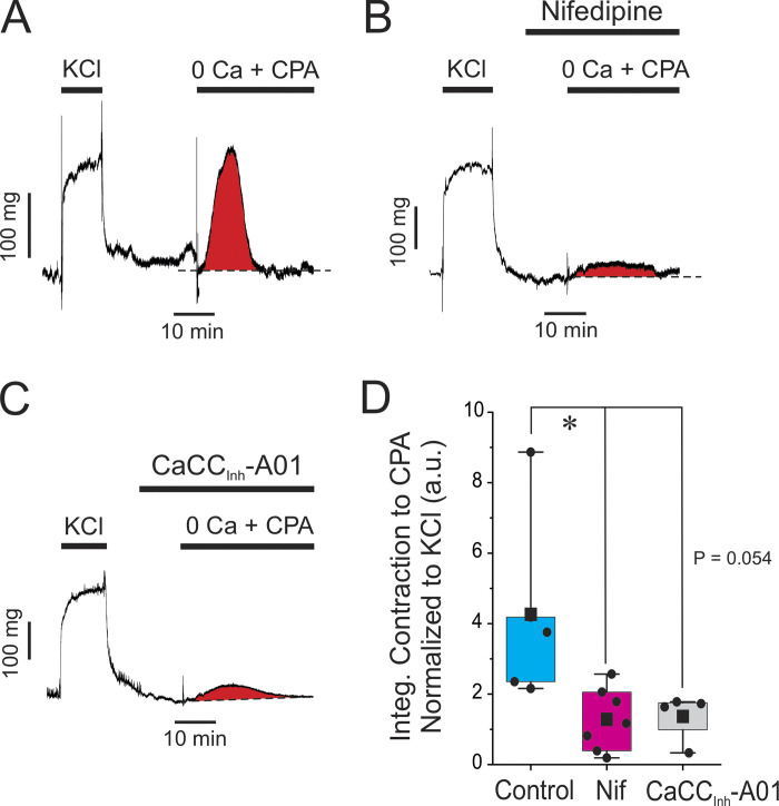
Blocking ANO1 or CaV1.2 depletes SR Ca2+ stores. (A) Typical isometric force recording obtained under control conditions showing the effect of depleting the SR Ca2+ stores. After eliciting a sustained contraction with high K+ (KCl; 85.4 mM), the preparation was switched to normal Krebs for 20 min, which relaxed the artery to baseline. The solution was subsequently changed to a Ca2+-free Krebs solution containing 100 μM EGTA and 10 μM cyclopiazonic acid (0 Ca + CPA), which triggered a transient contraction produced by releasing Ca2+ from internal SR Ca2+ stores. The area under the curve for the transient contraction highlighted in red was measured and used as an index of the amount of Ca2+ stored in the SR. (B and C) Similar experiments to that shown in A, with the exception that each preparation was incubated for 10 min with 1 μM nifedipine or 10 μM CaCCInh-A01, respectively, prior to switching to the Ca2+-free solution with CPA in the presence of either drug. Both compounds exerted a strong inhibition of the transient contraction elicited by CPA as evident from the much reduced area under the curve shown in red. (D) Pooled data from similar experiments are shown in A–C. For each dataset, the mean is indicated by a filled black square with the colored boxes and whiskers delimiting the 25th and 75th percentile, and the 10th and 90th percentile of the pooled data, respectively, and small dots individual data points of the integrated contraction measured in the presence of 0 Ca + CPA, normalized to the KCl-induced contraction (a.u.: arbitrary unit). Control, N = 5; nifedipine, N = 7; CaCCInh-A01, N = 4; * indicates a significant difference between means with P < 0.05. The comparison between the Control and CaCCInh-A01 groups was just at the limit of significance as shown.

