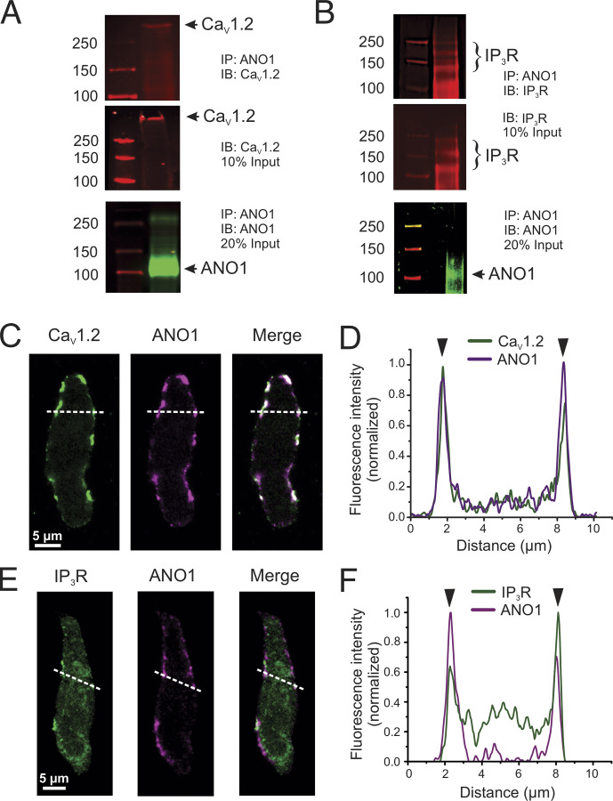
Figure 8.
ANO1, CaV1.2, and IP3R colocalize in peripheral coupling sites to form signaling complexes. (A and B) Co-IP of CaV1.2 or IP3R with ANO1 from lysates of the pulmonary artery from wild-type mice. Pulldown was carried out with anti-ANO1 antibody and then probed by Western blot with anti-CaV1.2, anti-IP3R, or anti-ANO1 antibodies. Five to six mouse tissues per experiment, each ran in triplicates. (C and D) Freshly isolated PASMCs from wild-type mice were immunolabeled for ANO1 and CaV1.2 (C) or ANO1 and IP3R (D). All three proteins were preferentially localized to the periphery of the cells. (D and F) Line profiles of the areas indicated by the white dashed lines in C and E. The fluorescence intensity was normalized to the minimum and maximum fluorescence for each sample. The black arrowheads denote the location of the PM. ANO1 and CaV1.2 show strong immunolabeling at the PM (D). (E) IP3R shows some intracellular immunolabeling, with moderate peaks present at the periphery showing an enhancement of protein localization to peripheral coupling sites. Source data are available for this figure: SourceData F8.

