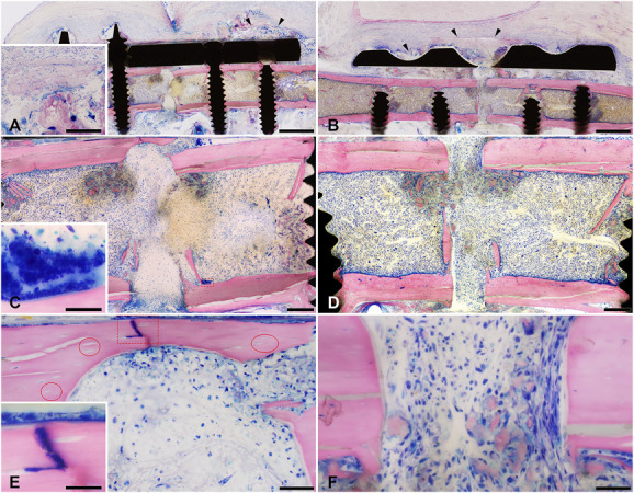
Fig. 6.

This figure shows representative microscopic images of Giemsa eosin-stained methyl methacrylate–embedded mouse femoral sections of Hi-SA5458-infected animals and Lo-SA5464-infected animals 4 days postoperatively (bone is stained pink, connective and soft tissue are stained rose, and cell nuclei are dark blue; the fracture site is either complete or only its cis or trans part is shown). Marked tissue necrosis was visible in (A, black arrow heads) Hi-SA5458-infected and (B, black arrow heads) Lo-SA5464-infected animals. Giemsa-positive coccoid bacteria were visible in (C, red square, inset) Hi-SA5458-infected animals. The straight cutting lines show the absence of new bone formation at the bone stumps in (C) Hi-SA5458-infected and (D) Lo-SA5464-infected animals. In (E) Hi-SA5458-infected animals (red circles), empty lacunae indicating osteonecrosis, the presence of a mainly polymorphonuclear or granulocytic inflammation accompanied by (E, red square, inset) Giemsa-positive coccoid bacteria were observed. Osteonecrosis was less pronounced in (F) animals infected with Lo-SA5464 and the inflammation was more mononuclear (no Giemsa-positive coccoid bacteria in this field of view). Artefacts: (D) There was black debris from the Gigli saw used to create the osteotomy at the initial surgery. (A, B) Cyan blue–stained, round structures in the soft tissue were monofilament sutures placed postmortem to fix the soft tissue to avoid shrinkage artefacts. Images were taken at 2 x (A and B: scale bar 1 mm), 7.5 x (C and D: scale bar 200 mm), 40 x (E and F: scale bar 50 mm), and 100 x oil (insets: scale bar 20 mm). A color image accompanies the online version of this article.
