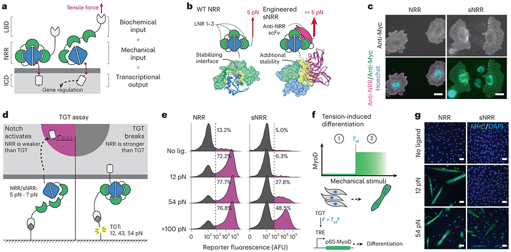
Fig. 1: Design of customized mechanosensation.

a, Schematic of tension-mediated activation in Notch or SynNotch receptors. Application of sufficient tensile force via the LBD activates the receptor by displacing three LNR modules and converting the NRR into a substrate for proteolysis at S2. Cleavage at S2, and subsequently at S3, liberates the ICD (LNR modules, green; S2 and S3, red). b, Activating the Notch1 NRR (left) requires tensile force to disrupt the intramolecular interactions that promote an autoinhibited conformation. Engineered sNRR domains (right) include an intramolecularly bound scFv for additional stability. The heterodimerization domain (HD) and LNRs of the NRR are blue and green, respectively, and the scFv added in sNRR is magenta. Molecular interactions between the LNRs and HD (left) or scFv and NRR (right) are yellow. (Protein Data Bank (PDB) codes: 3ETO and 3L95, respectively). c, Representative immunostaining of NRR- and sNRR-based receptors for surface-receptor with anti-Myc (green) and available NRR with exogenous soluble anti-NRR scFv (magenta) on HeLa cells. Scale bar is 25 μm. d, Schematic of TGT assay used to evaluate molecular tension needed to activate engineered receptors. FITC is used as a ligand for SynNotch receptors expressing an anti-FITC scFv LBD. e, NRR- or sNRR-based receptors with a Gal4-VP64 ICD induce expression of a UAS-controlled H2B-mCherry reporter upon activation. HEK293-FT cells transiently expressing these receptors were cultured on TGT-coated surfaces, and reporter activity was monitored by flow cytometry. Traces represent normalized densities of the total cells from three independently transfected pools (n = 3) expressing a cotransfection marker based on the mTurquoise2 within the analyzed populations. f, Tension-induced myogenic differentiation. C3H/10T1/2 fibroblasts express NRR- or sNRR-based SynNotch receptors with a TetR-based/tTA ICD. Upon activation, these receptors drive expression of p65-MyoD, which in turn leads to differentiation. g, Representative immunostaining identifies differentiation into skeletal myocytes by multinucleation and positive myosin heavy chain expression (green). Scale bar is 100 μm. LNR, LIN12-Notch repeat.
