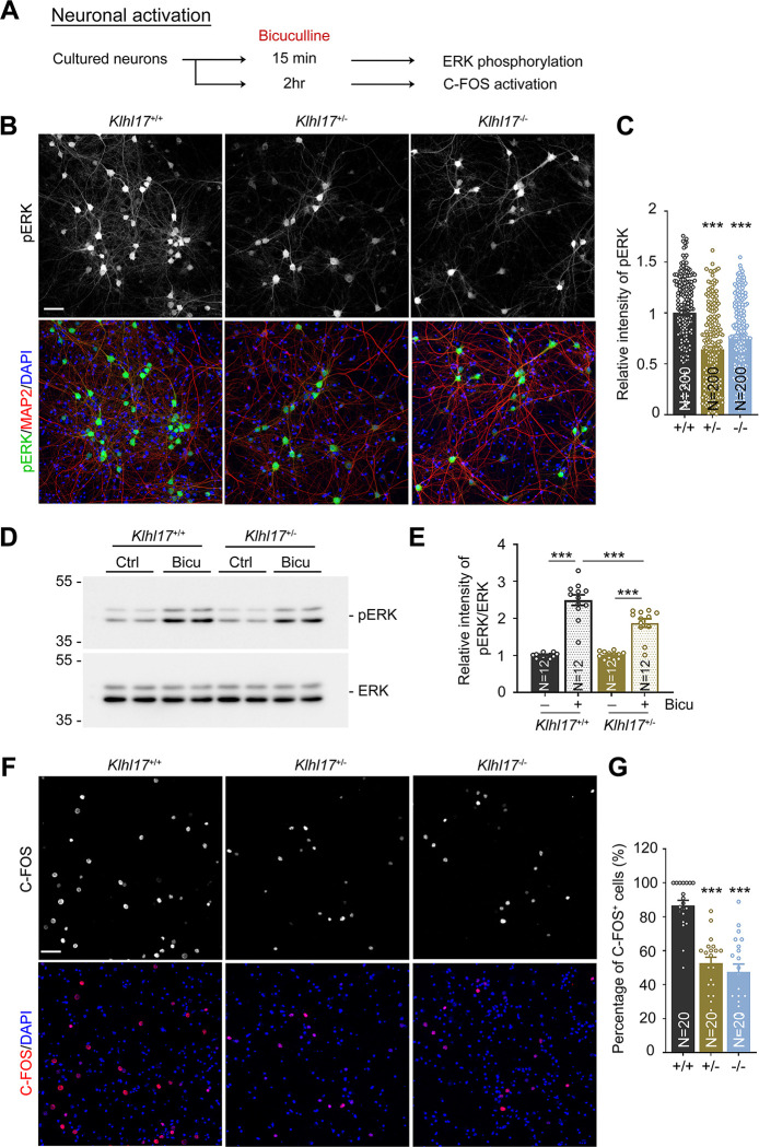
Fig 3. Klhl17 deficiency reduces ERK activation and C-FOS expression upon bicuculline treatment.
(A) Experimental flowchart. Cultured neurons from different genotypes were treated with bicuculline (40 μM) for 15 min to induce ERK phosphorylation (B–E) or 2 h to trigger expression of the immediate early gene C-FOS (F, G) at 18 DIV. (B–E) Both Klhl17+/–and Klhl17–/–neurons exhibit reduced ERK phosphorylation upon bicuculline treatment. (B) Representative images of immunostaining. Cultured neurons were stained with the neuronal marker MAP2 and the nuclear marker DAPI. (C) Quantification of the relative intensity of phospho-ERK signals based on immunostaining. (D) Immunoblots of ERK phosphorylation. (E) The quantification of ERK phosphorylation based on immunoblotting. (F, G) Numbers of C-FOS-positive cells induced by bicuculline are diminished upon Klhl17 knockout. (F) Representative images. Cultured neurons were stained with C-FOS antibody and DAPI. (G) The percentage of C-FOS-positive neurons. Samples were randomly assigned to treatments and collected from 2 independent experiments. The sample size (N) indicates the number of analyzed neurons in (C), the number of independent preparations in (E), and the number of image fields in (G). The data represent mean ± SEM. Individual data points are also shown. *** P < 0.001; one-way ANOVA. Scale bars: (B, F) 50 μm. The numerical value data and statistical results are available in S1 and S2 Data, respectively. DIV, day in vitro; ERK, extracellular signal-regulated kinase; KLHL17, Kelch-like protein 17.

