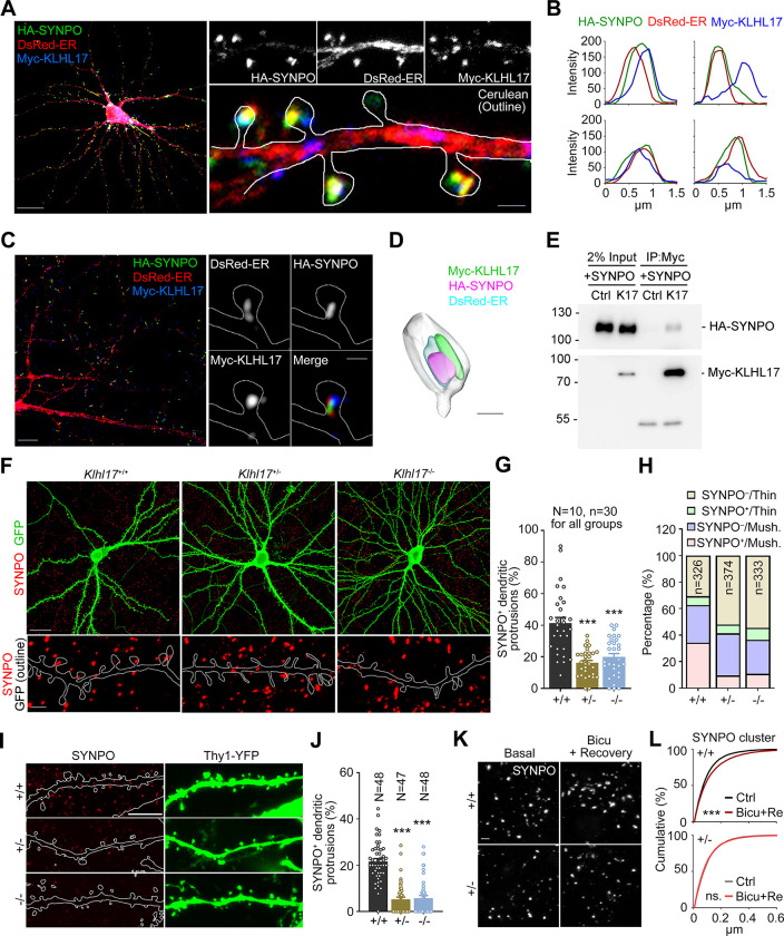
Fig 6. Klhl17 deficiency impairs synaptic expression and the distribution of synaptopodin.
(A–D) Colocalization or adjacent distributions of SYNPO and KLHL17 in cultured neurons. Cultured neurons were co-transfected with Myc-KLHL17, DsRed-ER, HA-SYNPO, and Cerulean constructs at 12 DIV and immunostained at 18 DIV. (A) Confocal images of the whole cell and enlarged dendrite segments. (B) Line scan results for Myc-KLHL17, DsRed-ER, and HA-SYNPO in the dendritic spines. (C) Super-resolution images of the whole cell and enlarged dendritic spines. (D) 3D reconstruction image using Imaris. (E) Coimmunoprecipitation of KLHL17 and SYNPO in Neuro-2A neuroblastoma cells. Neuro-2A cells were cotransfected with Myc-KLHL17 and HA-SYNPO constructs and subjected to immunoprecipitation and immunoblotting using the indicated antibodies. (F–H) Klhl17-deficient neurons in culture have fewer SYNPO-positive dendritic spines. (F) Representative images of the whole cells and enlarged dendrite segments. (G) Quantification of (F), i.e., the percentage of SYNPO+ dendritic spines. (H) Quantification of the percentage of SYNPO+ protrusions in thin and mushroom-like (mush.) spines of Klhl17+/+, Klhl17+/–, and Klhl17–/–neurons. (I, J) The percentage of SYNPO-positive dendritic spines in vivo is also reduced by Klhl17 deficiency. CA1 neurons of mice expressing YFP driven by the Thy1 promoter were analyzed. (I) Representative images. (J) The percentage of SYNPO+ protrusions. (K, L) Activity-dependent SYNPO clustering. Cultured neurons were treated with bicuculline (40 μM) for 15 min and then underwent washout for recovery for a further 30 min, which altered activity-dependent SYNPO clustering. (K) Representative images of endogenous SYNPO immunoreactivity. (L) Quantification of the size of SYNPO puncta upon synaptic stimulation. Samples were randomly assigned to treatments and collected from 2 independent experiments. In (G) and (J), the sample size “N” indicates the number of examined neurons. In (G), “n” represents the number of examined dendritic segments. In (H), “n” represents the number of examined dendritic spines. In (L), 10 image fields for each group were randomly collected from 2 independent experiments. All SYNPO-positive puncta (Klhl17+/+/Ctrl: 6523; Klhl17+/+/Bicu+Re: 7680; Klhl17+/–/Ctrl: 5354; Klhl17+/–/Bicu+Re: 6672) in the images were analyzed. The data represent mean ± SEM. Individual data points are also shown. *** P < 0.001; ns, not significant. One-way ANOVA (G, J); Kolmogorov–Smirnov test for cumulative probability (L). Scale bars: (A) whole cells: 20 μm; enlarged segment: 1 μm; (C) whole cells: 5 μm; enlarged segment: 0.5 μm; (D) 0.5 μm; (F) whole cells: 20 μm; enlarged segment: 2 μm; (I) 5 μm; (K) 2 μm. The numerical value data and statistical results are available in S1 and S2 Data, respectively. DIV, day in vitro; KLHL17, Kelch-like protein 17.

