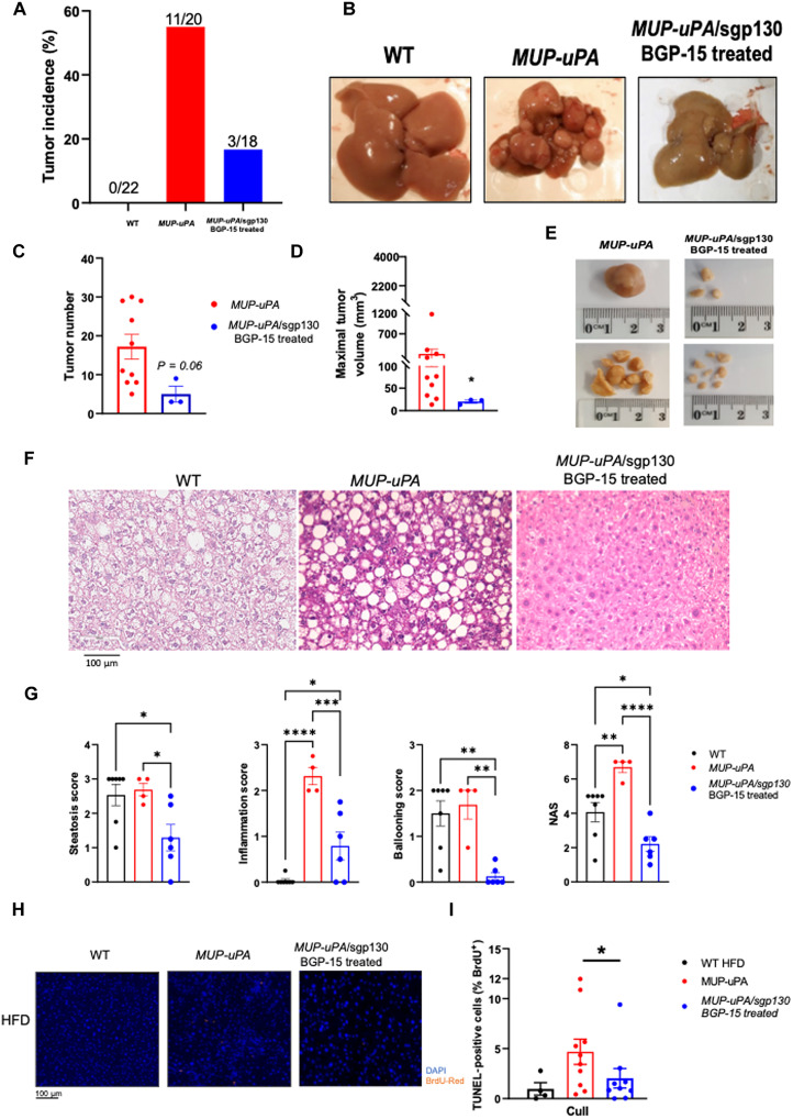
Fig. 3. Treatment of MUP-uPA/sgp130Fc mice with BGP-15 ameliorates NASHdriven HCC.
- Liver samples were obtained at 40 weeks. Tumor incidence in WT mice, MUP-uPA mice, and MUP-uPA/sgp130Fc mice treated with BGP-15 (A). Representative liver images (B). Tumor number (C), maximal tumor volume (D), and representative images (E). Representative H&E staining in WT, MUP-uPA, and MUP-uPA/sgp130 BGP-15–treated mice (F). Quantification of average lipid steatosis, lobular inflammation, hepatocyte ballooning, and NAS (G). The NAS is a measure of grade and is the sum of numerical scores applied to steatosis, hepatocellular ballooning, and lobular inflammation. Representative images (H) and quantification (I) for TUNEL staining. (C) and (D) were analyzed by unpaired t tests. (G) to (I) were analyzed by one-way ANOVA. The following numbers of biological replicates were used (independent mice) per group in each experiment: (A) 18 to 22; (C and D) 3 to 10; (F and G) 4 to 10. Data are expressed as means ± SEM. *P < 0.05, **P < 0.01, ***P < 0.001, ****P < 0.0001.

