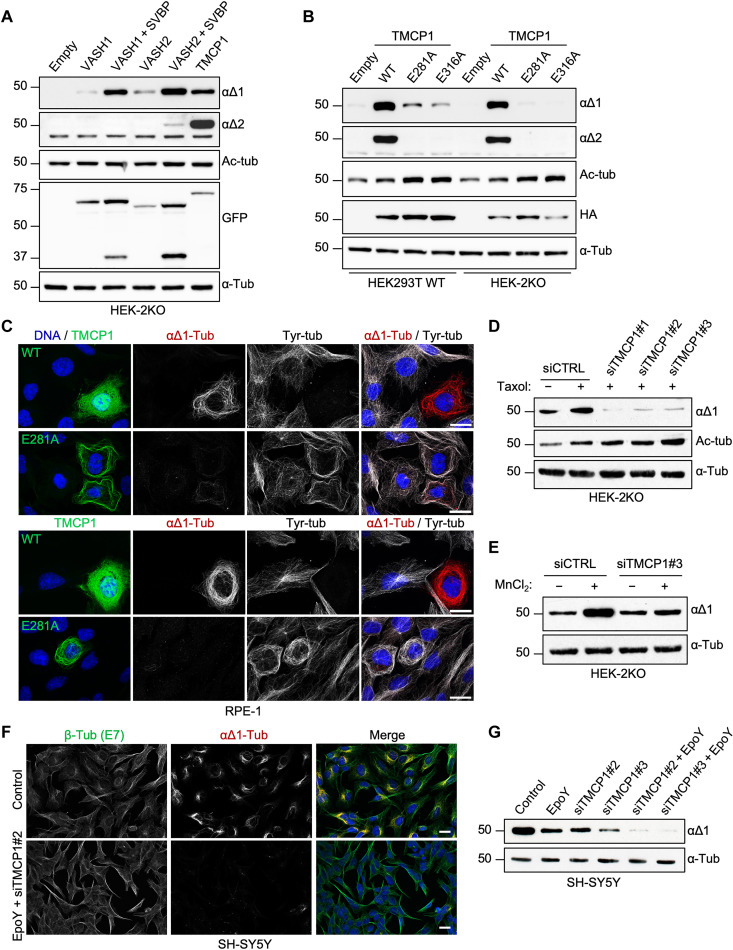
Fig. 2. TMCP1 catalyzes αΔ1- and αΔ2-tubulin.
(A) Immunoblots of protein extracts from HEK-2KO cells overexpressing VASH1 and VASH2, either alone or in combination with SVBP, and TMCP1. (B) Immunoblots of protein extracts from HEK293T or HEK-2KO cells expressing either the WT or enzymatically inactive mutants (E281A and E316A) of HA-TMCP1. (C) Immunofluorescence analysis of RPE1 cells expressing either active or inactive (E281A) GFP-TMCP1. Note the apparent MT bundling specific to cells expressing inactive GFP-TMCP1. Scale bars, 20 μm. (D) Immunoblots of protein extracts from HEK-2KO cells treated with paclitaxel following knock down of TMCP1 using three different siRNAs. (E) Immunoblots of an in vitro assay measuring tubulin detyrosination activity in control and TMCP1-depleted HEK-2KO cells. (F) Immunofluorescence analysis of SH-SY5Y cell knockdown for TMCP1 and simultaneously treated with the VASH inhibitor EpoY. (G) Immunoblots of protein extracts from SH-SY5Y cells treated with the EpoY inhibitor, following the knockdown of TMCP1 or after combination of the two treatments.

