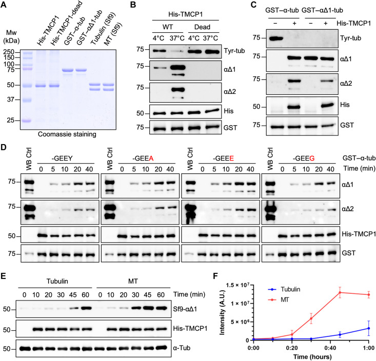
Fig. 3. Enzymatic mechanism and substrate specificity of TMCP1.
(A) Coomassie staining of purified proteins used for in vitro studies of TMCP1. His-TMCP1, His-TMCP1-E281A, GST–α-tubulin, and GST–αΔ1-tubulin were expressed in BL21 bacteria, whereas tubulin and MT were purified and assembled from insect (Sf9) cells. Mw, molecular weight. (B) Immunoblots of an in vitro assay using recombinant His-TMCP1 or its catalytically inactive version (E281A) and bacterially produced GST–α-tubulin. (C) Immunoblots of an in vitro assay involving bacterially produced GST–α-tubulin or GST–αΔ1-tubulin treated with recombinant His-TMCP1. (D) Immunoblots of an in vitro time-course assay using recombinant His-TMCP1 and either the WT GST–α-tubulin or its mutated versions in which the C-terminal tyrosine has been replaced by alanine, glutamate, or glycine. WB, Western blot. (E) Immunoblots of an in vitro assay measuring time-dependent activity of recombinant His-TMCP1 toward Sf9-derived tubulin dimers or MTs. (F) Graphical representation of His-TMCP1 activity toward Sf9-derived tubulin or MTs. Immunoblot signals were quantified for each time point (mean ± SD; n = 3 independent experiments). A.U., arbitrary units.

