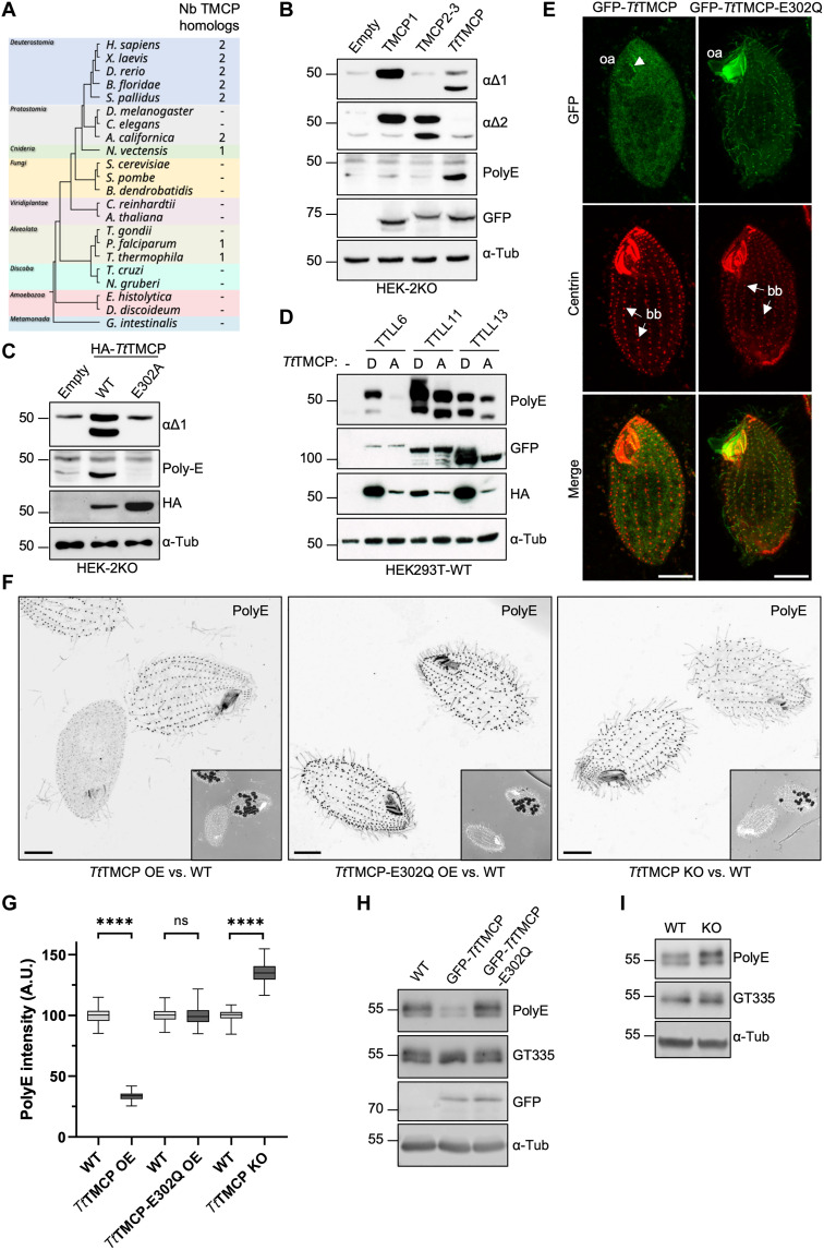
Fig. 7. Molecular and functional analysis of a TMCP homolog from Tetrahymena thermophila.
(A) Taxonomic tree of selected eukaryotes depicting the conservation of TMCPs. (B) Immunoblots of protein extracts from HEK-2KO cells expressing either GFP-TMCP1, GFP-TMCP2-3, or GFP-TtTMCP (Tetrahymena thermophila TMCP). (C) Immunoblots of protein extracts from HEK-2KO cells expressing TtTMCP or its enzymatically inactive version (E302A). (D) Immunoblots of protein extracts prepared from HEK293 cells coexpressing GFP-TTLL6, GFP-TTLL11, or GFP-TTLL13 together with either active or enzymatically dead HA-TtTMCP. (E) Immunofluorescence analysis of Tetrahymena cells showing the localization of GFP-TtTMCP or its enzymatically inactive version (E302Q) upon overexpression. Arrows are pointed at somatic basal bodies (bb), while the arrowhead indicates the basal bodies in the oral apparatus (oa). (F) Immunofluorescence analysis of Tetrahymena cells labeled with polyE antibody upon expression of either GFP-TtTMCP or GFP-TtTMCP-E302Q and the TtTMCP knockout cells. Comparison is made with WT cells on the same coverslip, which are identified by the presence of ink vacuoles. Scale bars, 10 μm. (G) Graphical representation of polyE signal intensity on the basal bodies from the experiments represented in (F) (****P < 0.0001 calculated with nested t test). ns, not significant. (H) Immunoblots of protein extracts from Tetrahymena cells expressing GFP-TtTMCP or its enzymatically inactive version E302Q as compared to WT. (I) Immunoblots of protein extracts from Tetrahymena cells knockout for TtTMCP as compared to WT.

