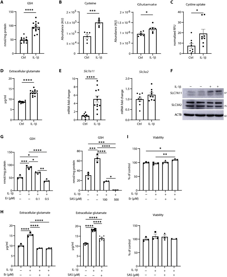
Fig. 1. IL-1β increases GSH and SLC7A11 levels in primary airway epithelial cells.
(A) GSH levels in control or IL-1β–stimulated mouse tracheal epithelial (MTE) cells following 24 hours of incubation with of IL-1β (10 ng/ml). (B) Levels of intracellular cysteine (left) and glutamate (right) in control and IL-1β–stimulated cells. AU, arbitrary units. (C) Fluorescein isothiocyanate–cystine uptake in control or IL-1β–treated cells. Results are expressed as relative fluorescence units (RFU) normalized to Hoechst staining in each well. (D) Levels of glutamate in conditioned medium from MTE cells in response to IL-1β. (E) Assessment of Slc7a11 and Slc3a2 mRNA. Ctrl, control. (F) Western blot analysis of SLC7A11 and SLC3A2 levels in response to IL-1β. ACTB, loading control. (G) GSH measurement in cells exposed to increasing doses of erastin (Er) or sulfasalazine (SAS). Cells were pretreated for 1 hour, followed by IL-1β treatment for 24 hours. (H) Extracellular glutamate levels in conditioned medium of cells pretreated with 0.5 μM Er or 100 μM SAS for 1 hour before IL-1β stimulation for 24 hours. (I) Assessment of cell viability in cells treated with 0.5 μM Er or 500 μM SAS and vehicle/IL-1β. *P < 0.05, **P < 0.01, ***P < 0.001, and ****P < 0.0001.

