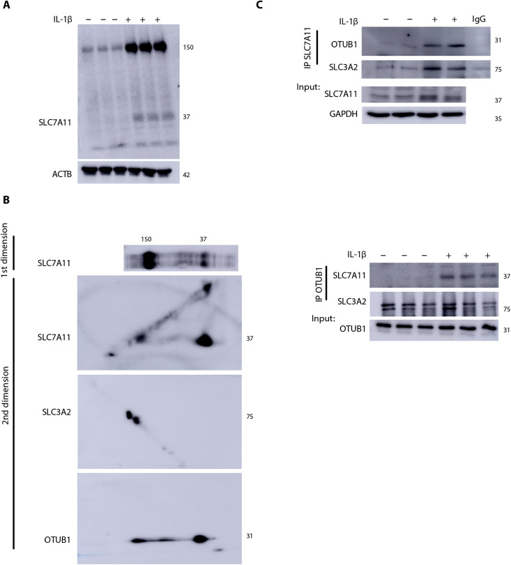
Fig. 2. Formation of an oxidation-dependent HMW complex of SLC7A11, SLC3A2, and OTUB1 in response to IL-1β.
(A) Nonreducing Western blot of SLC7A11 showing a high–molecular weight (HMW) complex around ~150 kDa in MTE cells that is further increased upon treatment with IL-1β as described in Fig. 1. ACTB, loading control. (B) Two-dimensional (2D) gel analysis of the nonreduced gel strip containing SLC7A11 (top). SLC7A11, SLC3A2, and OTUB1 were evaluated via Western blotting in the second dimension. (C) Top: Immunoprecipitation of SLC7A11 in MTE cells and Western blot analysis of OTUB1 and SLC3A2. SLC7A11 and glyceraldehyde-3-phosphate dehydrogenase (GAPDH) from whole-cell lysates were used as the input control. Bottom: Immunoprecipitation of OTUB1 protein followed by Western blotting analysis of SLC7A11 and SLC3A2. OTUB1 levels from whole-cell lysates were used as the input control. IgG, immunoglobulin G; IP, immunoprecipitation.

