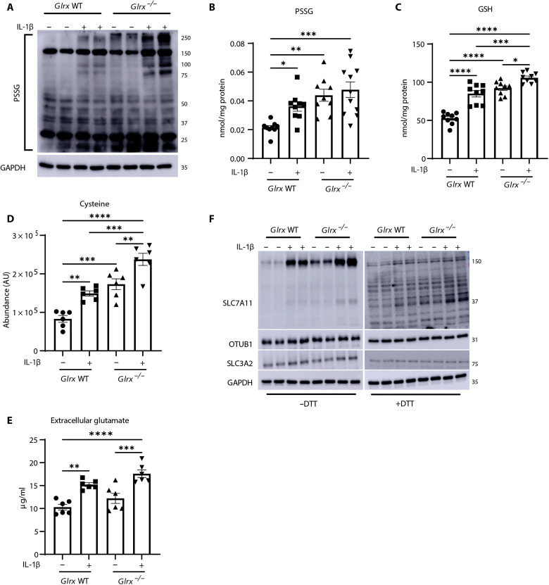
Fig. 3. S-glutathionylation regulates GSH in association with modulation of xC−.
(A) Assessment of PSSG in wild-type (WT) or Glrx−/− MTE cells treated with vehicle or IL-1β as described in Fig. 1. GAPDH, loading control. PSSG (B) or GSH (C) levels in WT or Glrx−/− epithelial cells stimulated with IL-1β for 24 hours. Intracellular cysteine (D) or glutamate content in conditioned medium (E) of WT or Glrx−/− epithelial cells stimulated with vehicle or IL-1β for 24 hours. (F) Nonreducing [−dithiothreitol (−DTT); left] and reducing (+DTT; right) Western blots of SLC7A11 (37 kDa), OTUB1, and SLC3A2 in WT and Glrx−/− cells. GAPDH, loading control. *P < 0.05, **P < 0.01, ***P < 0.001, and ****P < 0.0001.

