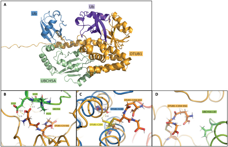
Fig. 8. Molecular modeling visualizing the impact of OTUB1-SSG on the binding between OTUB1 and UBCH5A.
(A) Protein complex of OTUB1 (orange), UBCH5A with Ub bound (green/blue), and the distal Ub (purple). Models were constructed with GSH at either C23 (OTUB1-C23-SSG, as shown) or C204. (B) In silico binding site between GSH-C23 (OTUB1-C23-SSG) (orange) and V120, P121, and E122 of UBCH5A (green). Contacts were shown to optimize over simulation length, with convergence of two sub–3-Å contacts between the glutamic acid of GSH and E122 of UBCH5A. (C) Visualization of OTUB1-C204-SSG disrupting minor polar contact between C204 and Ser180 within OTUB1. (D) Final frame of C204-SSG simulation showcasing minor nonpolar contact observed between GSH and L97 at UBCH5A interface.

