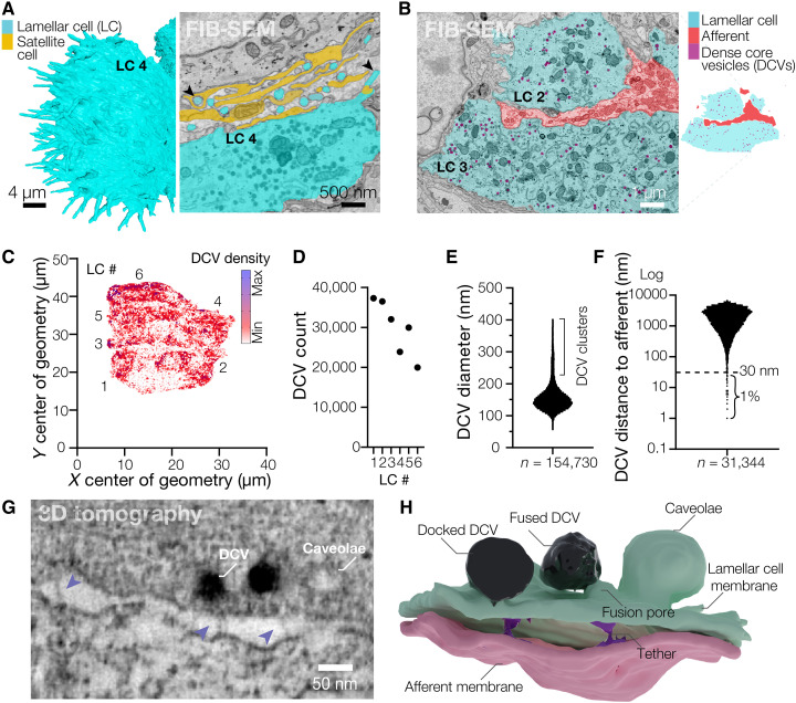
Fig. 2. Lamellar cells interact with the afferent via DCVs and tethers.
(A) Close-up 3D reconstruction of villi protruding from the edge of LC4 and a pseudo-colored scanning electron microscope image of villi (black arrowheads) protruding from the edge of LC4 and contacting the satellite cell and afferent. (B) A pseudo-colored scanning electron microscope image depicting DCVs inside LCs (left) and a corresponding map of the cell types shown in the image (right). (C) A density map of DCVs in LCs. (D to F) Quantification of the total DCV count per LC (D), DCV diameter (E), and distance from each DCV to the closest afferent membrane (F). n is the total number of DCVs. (G) Transmission electron microscopy image of the LC-afferent contact area. Blue arrowheads point to tethers connecting LC and afferent plasma membranes. (H) Three-dimensional reconstruction of a fragment of the LC-afferent contact area.

