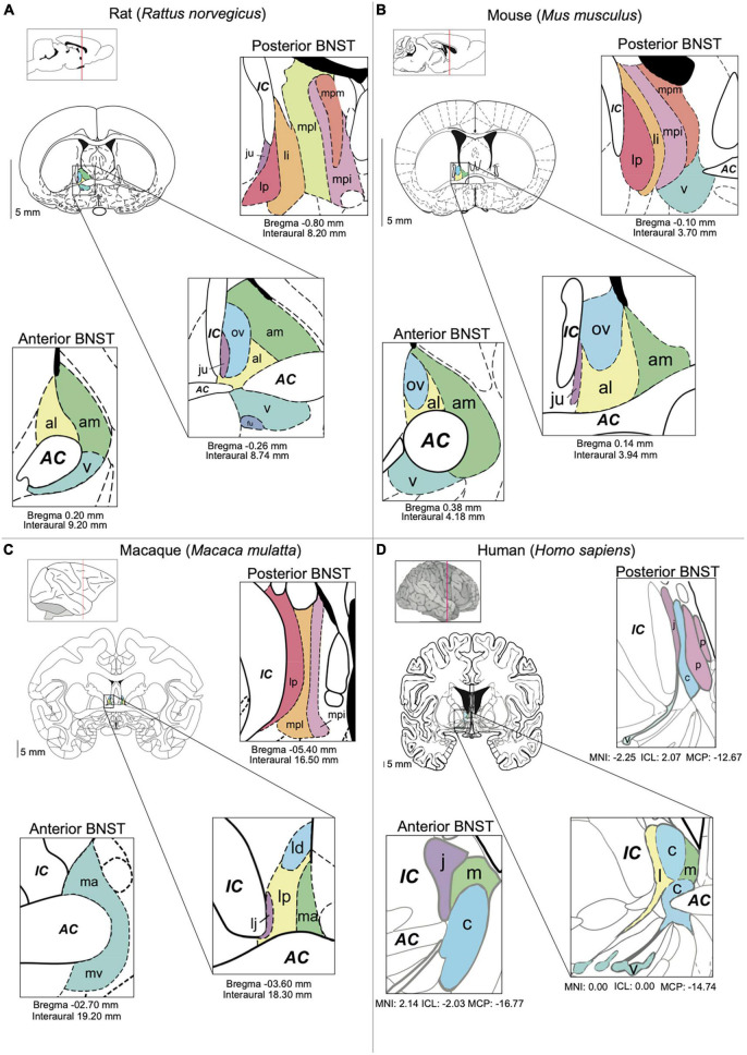
FIGURE 1.
BNST anatomy and nomenclature in different species. (A) Main nuclei of the BNST in the rat [Paxinos (1995) The Rat Brain in Stereotaxic Coordinates, 2nd edition]. (B) Main nuclei of the BNST in the mouse. (C) The BNST in the macaque (Paxinos et al., 2000). The Macaque Brain in Stereotaxic Coordinates, 2nd edn.). (D) Main nuclei of the BNST in the human [Mai (2016). Atlas of the Human Brain. 4th Ed.]. Subnuclei are colored based on their anatomic similarities. AC = anterior commissure, al = anterolateral BNST, am = anteromedial BSNT, c = central BNST, IC = internal capsule, ju = juxtacapsular BNST, l = lateral BNST, ld = laterodorsal BNST, lp = lateral posterior BNST, li = lateral intermediate BNST, m = medial BNST, ma = medial anterior BNST, mpi = medial posterointermediate BNST, mpl = medial posterolateral BNST, mpm = medial posteromedial BNST, mv = medioventral BNST, ov = oval BNST, p = posterior BNST, v = ventral BNST.

