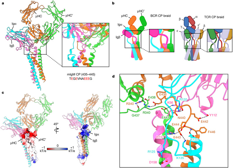
Fig. 3 ∣. CP interactions between the Igα/β heterodimer and mIgM.
a, Global view (left) and enlarged view (right) of the CP region of IgM BCR, showing the intertwining in this region. The sequence of mouse mIgM CP (435–445) is shown with acidic residues in red. b, Schematic diagrams for the CP assembly of BCR (left) and TCR (right). c, Electrostatic surfaces (−1 to +1 kT/e) at the CPs of mIgM (left) and the CPs of Igα and Igβ (right). d, Detailed interactions between the charged CP residues of the Igα/β heterodimer and mIgM.

