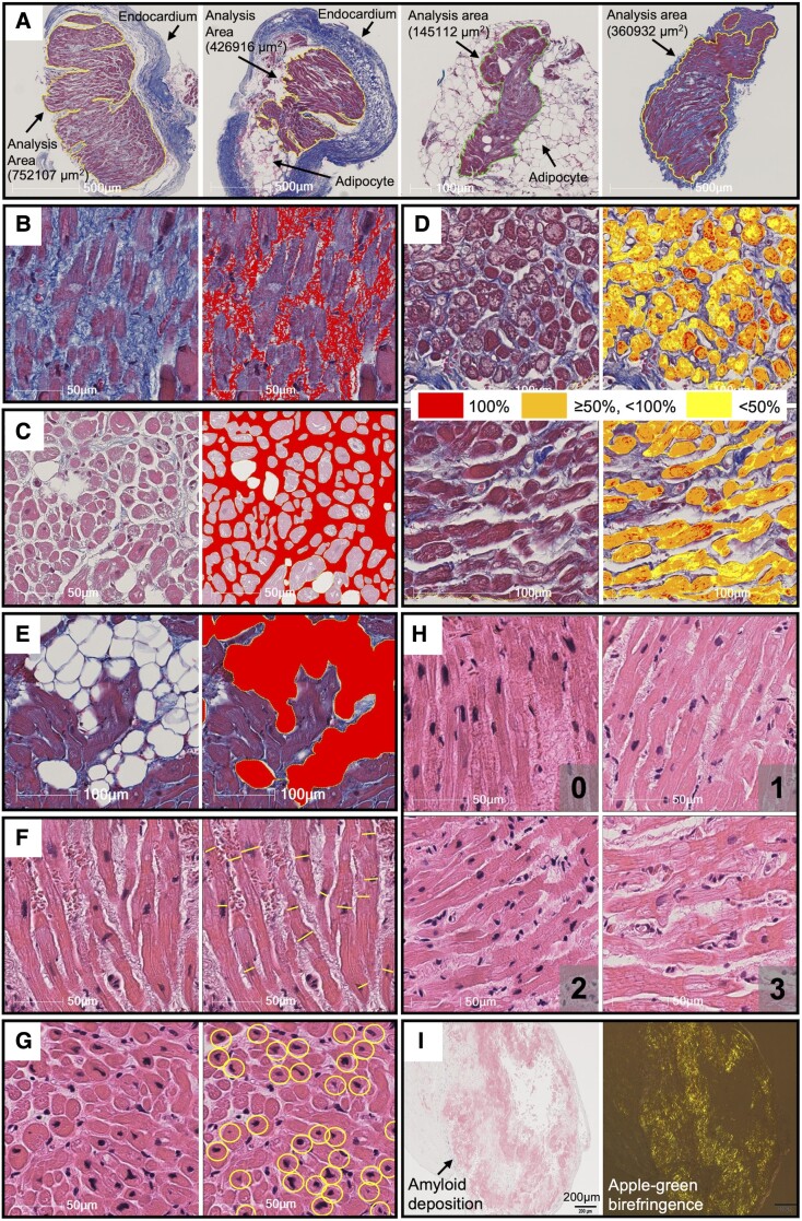
Figure 1.
Examples of measurement of histological factors. Analysis area was defined as the area surrounded by the solid lines excluding the endocardium and large adipose tissues (A). On Masson’s trichrome staining, fibrosis extent (B), intercellular space extent (C), myofibrillar loss severity (D), and adipocyte extent (E) were evaluated. On hematoxylin and eosin staining, myocyte size (F), and number of myocardial nuclei (G) were evaluated. Myocyte disarray was semi-quantitatively analysed and classified as minimal (0), mild (1), moderate (2), or severe (3) (H). Amyloid deposition was identified by Congo red staining and birefringence under polarizing microscope (I). See Supplementary data online, Appendix for the details.

