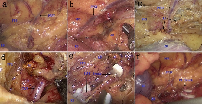
Figure 1.
Key procedures of the PFSV and CAV trunk during retroperitoneal laparoscopic left adrenalectomy and the main positional relationship of intraoperative anatomical structures to each other. (a) The PFSV trunk, the surface of the upper kidney, and the inner surface of PFS; (b) The lateral margin of golden AD is visible when the upper edge of PFS is incised; (c) The perirenal fat of AD was completely separated; (d) The positional relationship between CAV and the lower pole of AD; (e) The CAV was ligated with three Hem-o-lock clips; f The PFSV emanates from the left RV and travels upward into the direction of the diaphragm. PFSV perinephric fat sac vein, PFS perinephric fat sac, AD adrenal gland, CAV central adrenal vein, KD kidney, RA renal artery.

