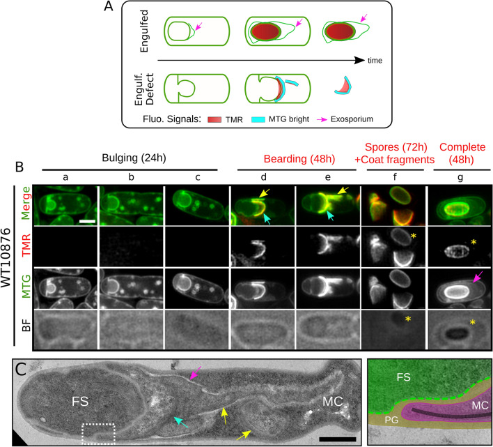
Figure 4.
Fluorescence SIM and TEM analysis reveal a coat bearding phenotype in engulfment-defective B. cereus cells. (A) Scheme of SIM localizations observed for the TMR-star (in red), the bright MTG signals (in cyan) and the MTG labelling the exosporium (pink arrows) in WT10876 cells presenting a complete engulfment (“Engulfed”) or an engulfment defect (Engulf. Defect). WT10876 cells sporulating at 20 °C in SMB were imaged with SIM and BF illumination modes after labeling with TMR-star and MTG (B) or by TEM after thin sectioning (C). (B) Typical sporangia showing an engulfment-defective phenotype detected at hour 24 and not presenting TMR-star or MTG bright signals (“bulging”, panels a-c) or at hour 48 and showing TMR-star and MTG signals (“bearding”, panels d-e) are shown. Sporangia with a refractile forespore detected at hour 48 (panel g, yellow asterisks) and spores (yellow asterisks) together with lysis fragments observed at hour 72 (panel f) are also shown for comparison. Cyan arrows show curved MTG bright signals close to the bulging membrane and yellow arrows point to MTG bright signals, which do not appear to be associated with the bulging membrane and drawing a straight line in the medial focal plan. Pink arrow points to the exosporium. Scale bar is 1 μm. (C) WT10876 cells sporulating at 20 °C, collected at hour 48 showing a blocked engulfment observed with TEM. Cyan arrow points to the large bearding coat fragment and yellow arrows point to the smaller fragments of coat disseminated in the MC cytoplasm. Pink arrow points at exosporium material. Right panel shows an inset (dotted white box on the left panel) of the junction between the FS (in green) and the MC (in magenta) compartments, separated by a thick peptidoglycan (PG) material (brown) with polymerized coat fragments (black dashed line) present until the leading edge of the MC membranes. Scale bar represents 0.6 μm.

