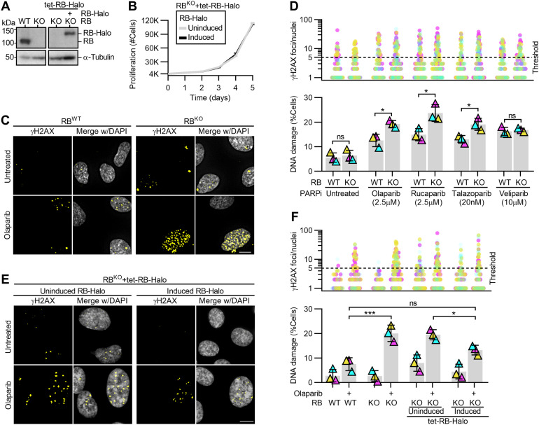
Figure 2. PARP trapping sensitizes RB-deficient cells to high levels of DNA damage.
(A) Western blot analysis of control (WT) and RB1-null (KO) RPE cells with and without doxycycline inducible RB-Halo (tet-RB-Halo). Cells were induced to express RB-Halo, as indicated (+). (B) Quantification of cell number in proliferative populations of RPE RBKO tet-RB-Halo cell number with and without 2 μg/ml doxycycline induction. (C, D) Representative images and quantification of γH2AX foci in RPE RBWT and RBKO cells with and without 48 h of incubation with the indicated PARP inhibitors. (E, F) Representative images and quantification of γH2AX foci in RPE RBKO tet-RB-Halo cells with and without doxycycline-induced RB-halo expression and after 48 h incubation with olaparib. (D, F) show the number of γH2AX foci per cell (top) and percent of cells with ≥5 damage foci (bottom). Scale bars are 10 μm. Experiments were performed and statistics calculated between independent experiments were performed in triplicate. Error bars represent SD between replicates. (*) P < 0.05; (**) P < 0.01; (ns) nonsignificant P > 0.05.

