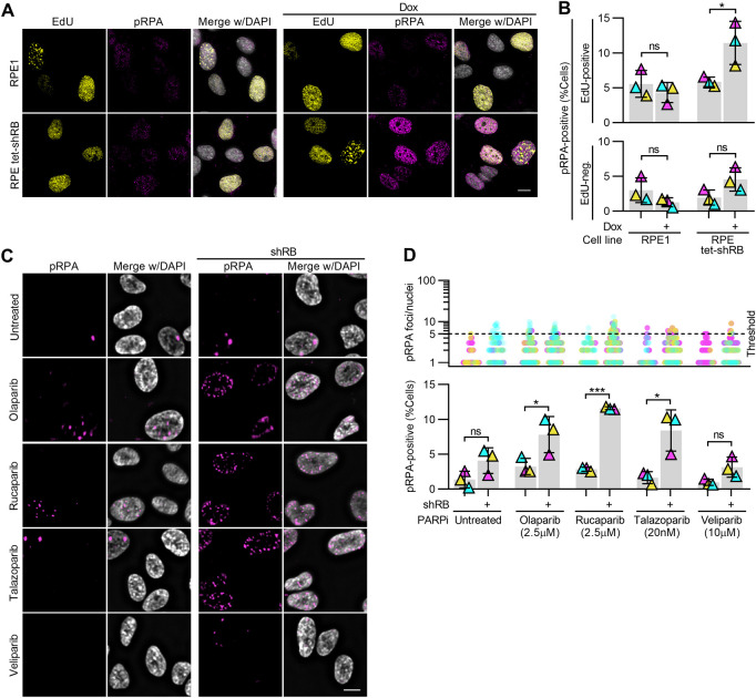
Figure S3. RB-depleted cells exhibit replication-dependent DNA damage after PARP trapping.
(A, B) Representative images and quantification of EdU and pRPA staining intensity in RPE1 and RPE tet-shRB cells with and without 2 μg/ml doxycycline as indicated (+). Thresholds for nuclear EdU and pRPA staining intensities were set on a per replicate basis and kept consistent across conditions within the replicate. (C, D) Representative images and quantification of pRPA foci in RPE tet-shRB cells with and without 2 μg/ml doxycycline induced shRB expression, after 48 h of incubation with PARP inhibitors, as indicated (+). (D) shows the number of pRPA foci per cell (top) and percent of cells with ≥5 foci (bottom). Scale bars are 10 μm. Error bars represent SD and statistics were performed between three independent experimental replicates. Data in (B) were analyzed using two-way ANOVA and Tukey’s multiple comparisons test. (*) P < 0.05; (***) P < 0.001; (ns) nonsignificant P > 0.05.

