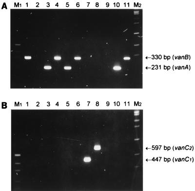
FIG. 1.
PCR analysis of VRE. Enterococci were subjected to PCR analysis, as described in Materials and Methods, with primers VanABF, VanAR, and VanBR (A) or VanC1F, VanC1R, VanC23F, and VanC23R (B). The PCR mixtures were electrophoresed on 2% agarose gels and stained with ethidium bromide. Lanes: 1, E. faecalis ATCC 51299; 2, E. faecium ATCC 19434; 3, E. faecalis 91; 4, E. faecalis 3; 5, E. faecium 143; 6, E. faecium 135; 7, E. gallinarum 129; 8, E. casseliflavus 38; 9, E. faecalis 26; 10, E. faecalis 21; 11, E. faecium 30. Lane M1, pUC19 DNA digested with HpaII (fragments of 501 and 489, 404, 331, 242, 190, and 147 bp are visible); lane M2, bacteriophage SPP1 DNA digested with EcoRI (fragments of 1,950, 1,860, 1,510, 1,390, 1,160, 980, 720, 480, and 360 bp are visible.

