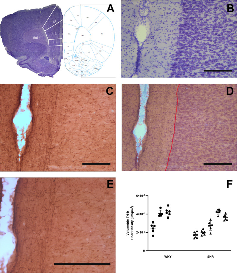
Fig. 4.
TH-ir fiber density in layer I of the PrL subregion of mPFC. A, B Coronal brain sections are used to identify the PrL of the mPFC (at around Bregma 3.00) on adjacent sections that are immunostained for TH (C, E). TH-ir fibers are encountered at all layers of the cortex, while those in Layer I are selectively oriented parallel to the pila surface (D, E). TH-ir fiber density measurements were confined in layer I, between the pial surface and Layers I and II border (red line). All scale bars = 250 µm. F The volumetric density of fibers in Layer I of the mPFC-PrL of SHR and WKY rats (n = 3 WKY and 5 SHR; 6 sections per animal; p > 0.05 in Nested t-test). PrL: prelimbic area; IL: infralimbic area; Cg1: anterior cingulate cortex

