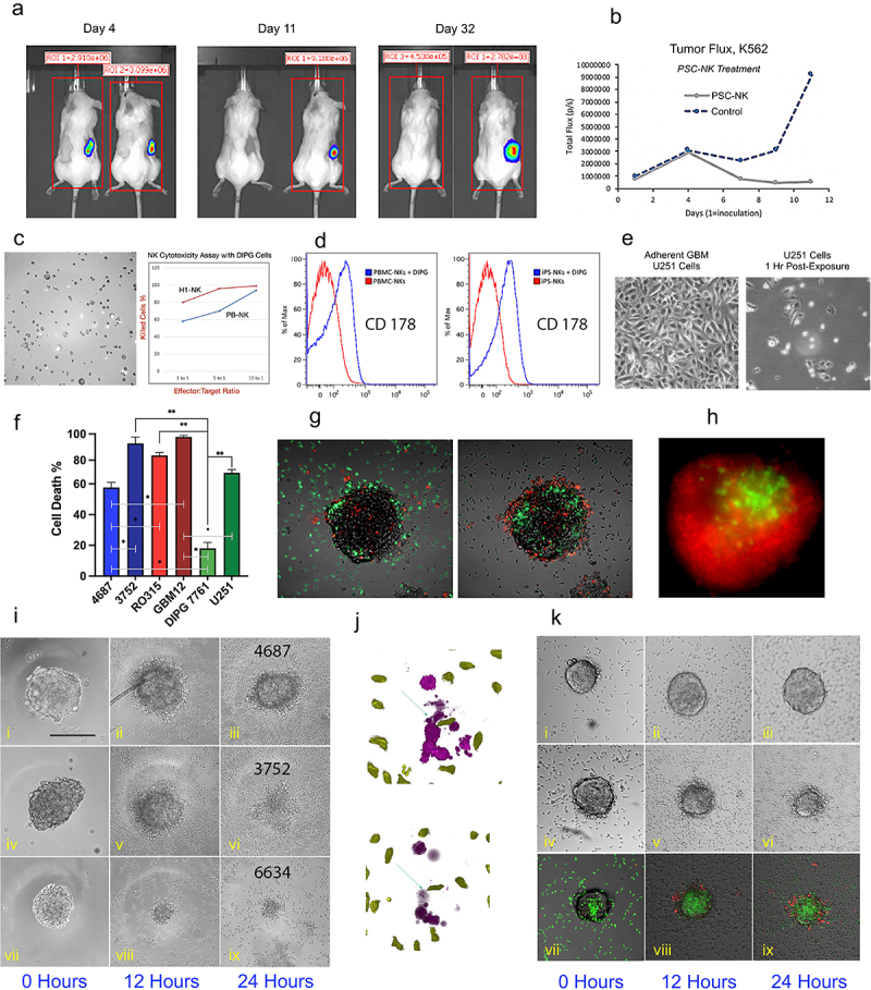
Figure 4.

hPSC-NK Cell Cytotoxicity Against Tumor Experiments.
a. Bioluminescence images showing chronic myelogenous leukemia tumor burden in two groups of mice on Day 4, Day 11, and Day 32 (post-tumor inoculation). hPSC-NK treatment was administered on Day 4 post-tumor injection. For each image, the treated group is on the left; the untreated control group is on the right. b. Tumor flux data collected from Day 1 (tumor inoculation) to Day 11 (post-inoculation). The graph is showing hindered tumor progression in mice receiving hPSC-NK cell treatment compared to control mice. c. PhC image and graph showing hPSC-NK cytotoxicity against DIPG cells. d. Flow cytometric analysis of FasL activation in NKs. Induction of FasL expression (CD178) was achieved via overnight incubation of NKs and DIPGs (SF8628). Both PB-NKs and hPSC-NKs responded to the DIPG stimulus, as indicated by the elevated cell numbers expressing FasL in comparison to untreated cells. e. PhC images showing hPSC-NK cytotoxicity against U251 GBM cells. f. A graph demonstrating hPSC-NK killing efficiency of various GBM cell lines. Bars represent mean ± SEM from at least three independent experiments. g. Overlay of PhC and immunofluorescence (IF) images demonstrating hPSC-NK (green) ability to penetrate the GBM tumor within 2 hrs of co-incubation. h. Fluorescence microscopy image showing hPSC-NK cytotoxicity against GBM. The GBM spheroid (6634) was eliminated (dead cells shown in red) by overnight exposure to hPSC-NKs (green). i. PhC images showing GBM neurospheres at 0 hrs, 12 hrs, and 24 hrs of incubation with hPSC-NKs; (i-iii) GBM4687 tumor cells are still present after 24 hrs of incubation with hPSC-NKs; (iv-vi) GBM3752 tumor cells are still present at the 12 hr incubation timepoint but were eliminated at the 24 hr timepoint; (vii-ix) GBM 6634 tumor cells are eliminated at the 12 hr incubation timepoint. j. Image showing hPSC-NKs (green) approaching tumor cells (purple, top) and causing tumor cell lysis (bottom). k. PhC and IF images showing hPSC-NKs co-incubated with GBM12 (with and without IL13Rα2 hAb); (i-iii) Without the addition of IL13Rα2 hAb, GBM6 inhibited hPSC-NK cell activity; (iv-vi) When IL13Rα2 antibody is added to the medium, hPSC-NKs eliminate GBM6 within 24 hrs of incubation; (vii-ix) hPSC-NKs are shown in green, and the apoptotic tumor cells are shown in red.
