FIGURE 1.

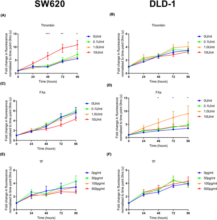
The effect of exogenous coagulation factors on proliferation in colorectal cancer cell lines. SW620 or DLD‐1 cells were seeded (5.0 × 10³ and 2.5 × 10³ cells per well, respectively). Exogenous clotting factors were added at 24 h (thrombin 0.0–10 U/mL, FXa 0.0–10 U/mL or TF 0–500 pg/mL). PrestoBlue® was added at intervals and fluorescence was measured. (A) Thrombin (10 U/mL) increased proliferation from 48 h onwards compared to the vehicle control in the SW620 cell line. There was no difference in proliferation at lower concentrations of thrombin. (C, E) There was no difference in proliferation in the SW620 cell line following the addition of FXa or TF at any concentration compared to the vehicle control. (B, F) There was no difference in proliferation in the DLD‐1 cell line following the addition of thrombin or TF at any concentration compared to the vehicle control. (D) FXa (1.0 U/mL) increased proliferation from 48 h onwards compared to the vehicle control in the DLD‐1 cell line. There was no difference at other concentrations of FXa. Data presented as mean fold change in fluorescence normalised to time point 0 h ±SEM. Data from at least three independent experiments. Statistical differences were determined using analysis of variance (ANOVA). *p < 0.05, **p < 0.01, ***p < 0.001. TF, tissue factor.
