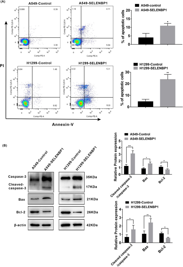
FIGURE 5.

Overexpression of SELENBP1 inducing the apoptosis of NSCLC cells under nonhigh level of oxidative stress was associated with the activation of caspase‐3 signaling pathway in vitro. (A) Flow cytometry analysis was conducted (left), and the ratio of apoptotic cells (%) was calculated (N = 3; right). All data were presented as the mean ± SD, unpaired t‐test, “*” p < 0.05, “**” p < 0.01, A549‐Control group versus A549‐SELENBP1 group and H1299‐Control group versus H1299‐SELENBP1 group. (B) The effect of SELENBP1 overexpressing on the expression of the components of caspase‐3 pathway was measured. A549‐SELENBP1, H1299‐SELENBP1, and their control cells lysate were collected and subjected to western blotting analysis with antibodies against the caspase‐3, cleaved‐caspase‐3, Bcl‐2, and Bax (left). Levels of β‐Actin were used as loading control. The histogram was used to quantify the experimental results of western blotting (N = 3; right). All data were presented as the mean ± SD, unpaired t‐test, “*” p < 0.05, “**” p < 0.01, A549‐Control group versus A549‐SELENBP1 group and H1299‐Control group versus H1299‐SELENBP1 group.
