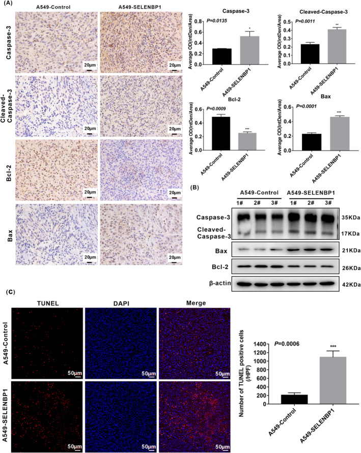
FIGURE 6.

Overexpression of SELENBP1 promoting the apoptosis of NSCLC cells in vivo was associated with the activation of caspase‐3 signaling pathway. (A) Paraffin sections of the retrieved tumor samples were subjected to IHC staining with antibodies against caspase‐3, cleaved‐caspase‐3, Bcl‐2, and Bax (left). Staining without primary antibody was used as negative controls. Results were observed under a bright field microscope (×400). Scale bars, 20 μm. The histogram was used to quantify the experimental results of IHC (N = 3; right). All data were presented as the mean ± SD, unpaired t‐test, “*” p < 0.05, “**” p < 0.01, “***” p < 0.005, A549‐Control tumors group versus A549‐SELENBP1 tumors group. (B) The expression of caspase‐3, cleaved‐caspase‐3, Bcl‐2, and Bax in mice tumor xenograft model were measured by western blotting (N = 3/group). Levels of β‐Actin were used as loading control. (C) The lever of apoptosis was presented by TUNEL staining (N = 3; left), and the histogram was used to quantify the TUNEL‐positive cells (right). Images were observed under a microscope (×200). Scale bars, 50 μm. TUNEL‐stained cells are in red, and DAPI‐stained nuclei are in blue. And the data were presented as the mean ± SD, unpaired t‐test, “***” p < 0.005, A549‐Control tumors group versus A549‐SELENBP1 tumors group.
